Nasal Fistulas
Surgery to close cerebrospinal fluid (CSF) and odontogenic fistulas
What are nasal fistulas
A nasal fistula is an abnormal communication, a kind of passage that should not exist, between the nasal cavity and a neighboring space that is normally separated from it. The name, however, brings together quite different situations. The two most important ones have opposite origins: one comes from above, from the compartment that surrounds the brain (the CSF fistula), and the other comes from below, from the mouth and the teeth (the odontogenic fistula, also called an oroantral communication). They have distinct causes, severity and treatment, and for that reason they deserve to be explained separately.
Two origins, two different conditions
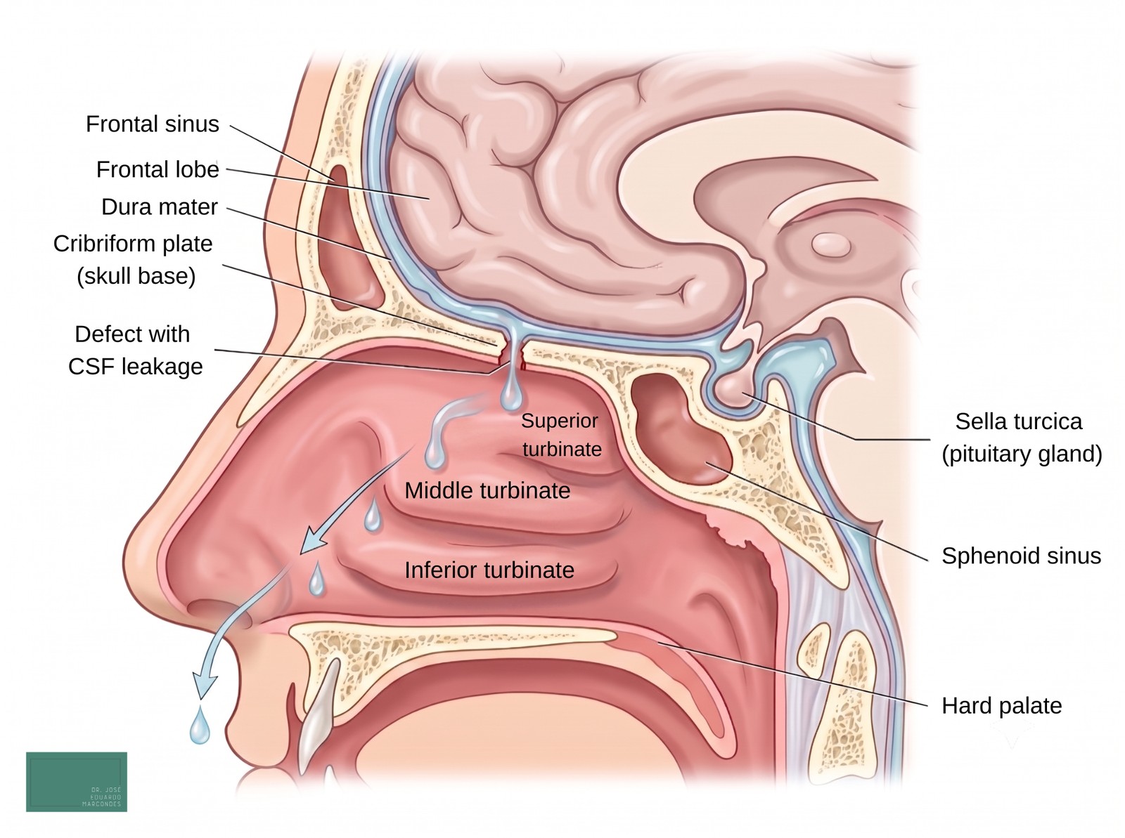
Understanding where the fistula comes from is the first step. In the CSF fistula, the defect is in the skull base, and the fluid that surrounds the brain (the cerebrospinal fluid) escapes into the nose. It is the most delicate one, because it opens a door for infections to reach the nervous system. In the odontogenic fistula, the communication is between the mouth and the maxillary sinus (the air cavity within the facial bone, behind the cheek), almost always after a problem in an upper tooth. It is less dangerous, but it usually causes recurrent sinusitis and discomfort. Below, each one in detail.
CSF fistula (CSF rhinorrhea)
What it is
The brain is bathed in a clear fluid called cerebrospinal fluid, contained within membranes and the skull. When a small defect appears in the bone and in the membrane (the dura mater) of the skull base, this fluid can leak into the nose. This leak is the CSF fistula, also called CSF rhinorrhea.
Why it is the most serious
The greatest risk is not the loss of fluid itself, but the pathway that the fistula opens: through this passage, bacteria from the nose can travel up and cause meningitis, a serious infection of the membranes of the brain. In persistent fistulas that are not treated, studies show a risk of meningitis of around 19%, with about 0.3 episodes per year while the fistula is active. For this reason, a confirmed CSF fistula requires specialized evaluation.
Causes
The most common cause is trauma, such as fractures of the face and the skull base. Next come the causes linked to surgery in the region (endoscopic sinus surgery, pituitary surgery, neurosurgery), called iatrogenic. There is also the spontaneous form, which appears without trauma and is associated with increased CSF pressure, idiopathic intracranial hypertension and obesity. Tumors and congenital defects complete the list. In classic series, about 80% of cases are traumatic, 16% are linked to surgery and 4% are spontaneous, a proportion that varies between centers, with a recent increase in the spontaneous forms.
Symptoms
The typical symptom is the dripping of a clear, watery fluid, usually from only one nostril, that worsens when the person lowers the head or strains. Many report a salty taste in the throat. Headache and, above all, repeated episodes of meningitis are important warning signs. A word of caution: the old “halo sign” (the ring that forms around blood on a tissue) is not reliable, because water and ordinary secretions also produce it. For this reason, confirmation depends on a laboratory test.
Diagnosis
The most accurate test to confirm that the fluid is indeed CSF is the measurement of beta-2-transferrin, a protein almost exclusive to CSF, with a sensitivity of around 94% to 100%. To locate the defect, high-resolution CT of the skull base is used and, when necessary, magnetic resonance imaging (MR cisternography). During surgery, a dye (fluorescein) can help identify the exact point of the leak.
Treatment
The treatment of choice is endoscopic endonasal repair, performed from inside the nose, without external cuts on the face. The surgeon closes the defect with grafts taken from the patient (such as fascia, mucosa or fat) and, when indicated, with vascularized flaps, such as the nasoseptal flap. The success rates reported in the literature are high: about 90% on the first attempt and around 97% after a second one, when needed. In the spontaneous form, treating only the defect is not enough: it is essential to control the CSF pressure, with weight loss, medication (such as acetazolamide) and, in selected cases, a shunt valve, because without this the fistula tends to recur. In specific low-flow situations, conservative measures (rest, head-of-bed elevation, a lumbar drain) can be tried.
Odontogenic fistula (oroantral communication)
What it is
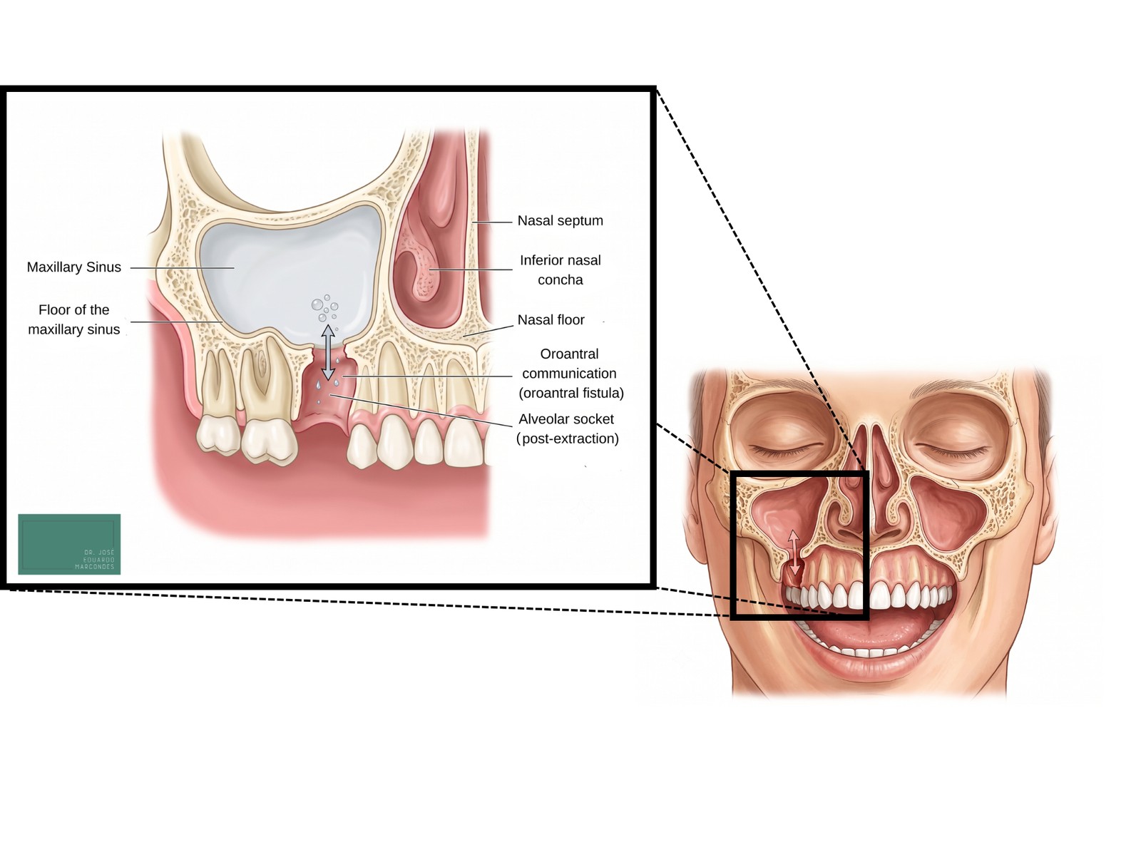
Here the communication is between the mouth and the maxillary sinus, the air-filled cavity in the facial bone, just above the upper back teeth. When this communication is recent, not yet lined, it is called an oroantral communication. When it becomes chronic and lined by mucosa, it becomes an oroantral fistula, which no longer closes on its own.
Causes
The most common cause is the extraction of upper back teeth, especially the first upper molar, whose roots lie very close to the floor of the maxillary sinus. Infections and abscesses of dental origin, cysts, implants and tumors can also cause the communication.
Symptoms
The most characteristic signs are the passage of liquids from the mouth into the nose (water comes back through the nose when drinking), air escaping through the mouth when blowing the nose, along with recurrent maxillary sinusitis, bad taste and bad breath. Some patients also notice a change in their speech.
Diagnosis
Diagnosis begins with the clinical examination, often with the maneuver of blowing with the nose held closed (Valsalva), which reveals the passage of air. CT confirms the defect, assesses the condition of the maxillary sinus and looks for root remnants or foreign bodies.
Treatment
Small and recent communications, usually less than 3 to 5 millimeters, can close on their own, as long as the infection is controlled and the clot is preserved. Larger or chronic fistulas, on the other hand, need surgical closure, with flaps from the mouth itself, such as the buccal advancement flap (Rehrmann technique), the palatal flap or the use of the buccal fat pad (Bichat’s fat pad). When there is associated maxillary sinusitis, it is treated at the same time, endoscopically or, in more extensive cases, through a sinus approach. The success rates of closure are high when the treatment is carried out properly and early.
When to seek evaluation
It is worth seeing an otolaryngologist (ENT) if you notice: a clear fluid dripping persistently from one nostril, especially after trauma or surgery; repeated episodes of meningitis; or the passage of liquids and air between the mouth and the nose after a dental extraction. The earlier the fistula is identified, the simpler the treatment usually is, and, in the case of the CSF fistula, the lower the risk of meningitis.
This content is informative and does not replace a medical consultation. Dr. José Eduardo Marcondes, ENT physician (CRM SP 107711 | RQE 43840).
Frequently asked questions about nasal fistulas
There is a clear fluid dripping from just one nostril. Could it be CSF?
It could be, especially if it is watery, comes out more when you lower your head or strain, and is accompanied by a salty taste, particularly after trauma or surgery. It cannot be confirmed by appearance alone: the test that clarifies it is the measurement of beta-2-transferrin in the fluid. Faced with this suspicion, seek evaluation, because a CSF fistula increases the risk of meningitis.
Is a nasal fistula dangerous?
It depends on the type. The CSF fistula, which comes from the compartment of the brain, is the most delicate, because it can open a path to meningitis. The odontogenic fistula, between the mouth and the maxillary sinus, tends to be less dangerous, but it causes recurrent sinusitis and discomfort, and it also needs to be treated.
Is CSF fistula surgery done from the outside, on the skull?
Most of the time, no. The repair is done from inside the nose, with an endoscope, without external cuts on the face, using grafts and flaps to close the defect. The success rates reported in the literature are high.
After having an upper tooth pulled, fluid started passing from the mouth to the nose. What is it?
This is a classic sign of an oroantral communication, an opening between the mouth and the maxillary sinus, which can happen after the extraction of upper back teeth. It should be evaluated promptly: small communications can close on their own with care, but larger or persistent ones need a procedure to close them.
Does every fistula need surgery?
No. Small and recent oroantral communications can heal on their own with infection control. A confirmed CSF fistula and persistent oral fistulas, on the other hand, usually need surgical closure. Management is always individual.