Hearing is the foundation of language, social interaction, and learning, and small changes in how a child hears can have a major impact on development. Identifying problems early, providing reassurance with reliable information, and acting promptly can reshape a child’s path in speech, school, and family life.
Why hearing matters from the very start
From birth, the brain learns language from the sounds in the environment and the voice of a caregiver. When hearing is not fully intact, a child may start speaking later, tire more easily in noisy settings, and rely on lip-reading to follow conversations, which can sometimes be mistaken for inattention or shyness.
Most common causes of hearing loss in children
– Otitis media with effusion: fluid behind the eardrum that reduces the passage of sound, common in early childhood and often silent.
– Congenital sensorineural hearing loss: it may be genetic, isolated, or part of a syndrome, and may remain stable or progress over time.
– Infections: cytomegalovirus during pregnancy and meningitis in childhood are among the most relevant infectious causes.
– Perinatal factors: prematurity, neonatal hospital stays, significant jaundice, and hypoxia increase risk and call for monitoring.
– Ototoxicity and noise: some medications and prolonged use of headphones at high volume may damage the cochlea, especially in school-age children and adolescents.
– Other conditions: earwax impaction, anatomical changes of the ear, and autoimmune diseases are also worth considering.
Signs that deserve attention
– In early life: little startle response to loud sounds, limited babbling, difficulty locating sounds, and a reduced response to their own name.
– In early childhood: delayed speech, persistent sound substitutions, a need to see the speaker’s mouth, and apparent inattention.
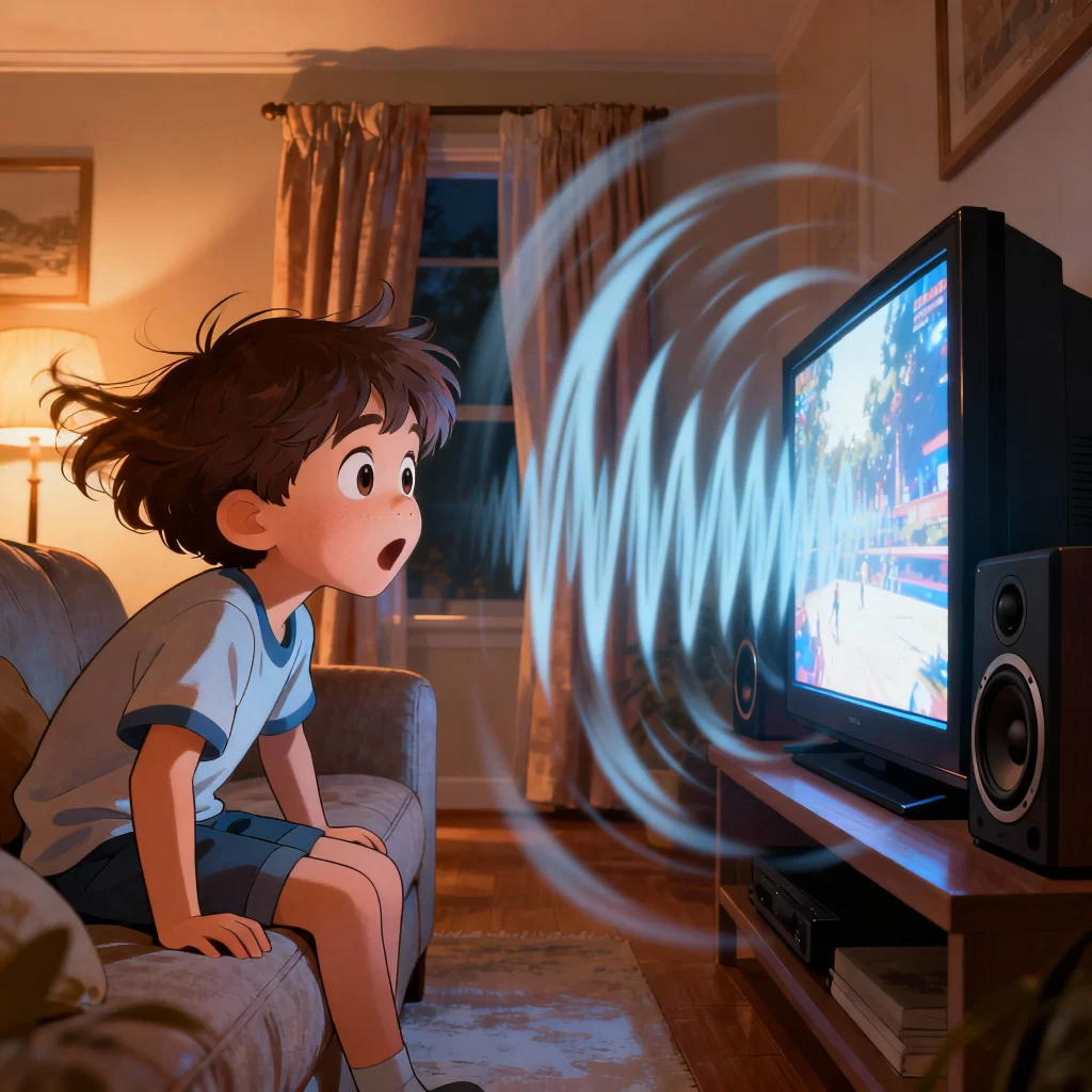
– At school age: keeping the television volume high, frequently asking for repetition, listening fatigue at the end of the day, and complaints of ringing in the ears (tinnitus).
– At any age: a family history of hearing loss, recurrent ear infections, rhinitis, and chronic mouth breathing are additional warning signs.
What to watch for day to day
– Language milestones: the progression from babbling to syllables, words, and sentences should occur steadily, even in bilingual settings.
– Noisy environments: disproportionate difficulty following conversations at parties, restaurants, and in the classroom may suggest mild to moderate hearing loss.
– Airway health: rhinitis, enlarged adenoids, snoring, and frequent ear infections warrant an otolaryngology evaluation.
– Exposures and history: headphones, neonatal hospitalization, marked jaundice, meningitis, and the use of medications with ototoxic potential indicate a need for closer follow-up.
How the diagnosis is made
– At the maternity ward: the newborn hearing screening with otoacoustic emissions is mandatory and can detect changes even before discharge, allowing for safe referral.
– The ideal timeline: screening by the first month, diagnostic confirmation by the third month, and the start of intervention by the sixth month take advantage of the best window of neuroplasticity.
– Objective and behavioral tests: otoacoustic emissions, auditory brainstem response, tympanometry, and play audiometry help define the type and degree of hearing loss accurately.
– Investigating the cause: genetic testing when indicated, imaging studies in selected cases, and testing for cytomegalovirus in the first weeks of life in suspected situations.
– Teamwork: integration between pediatric otolaryngology and speech-language therapy translates test results into a personalized treatment plan.
Treatment and rehabilitation
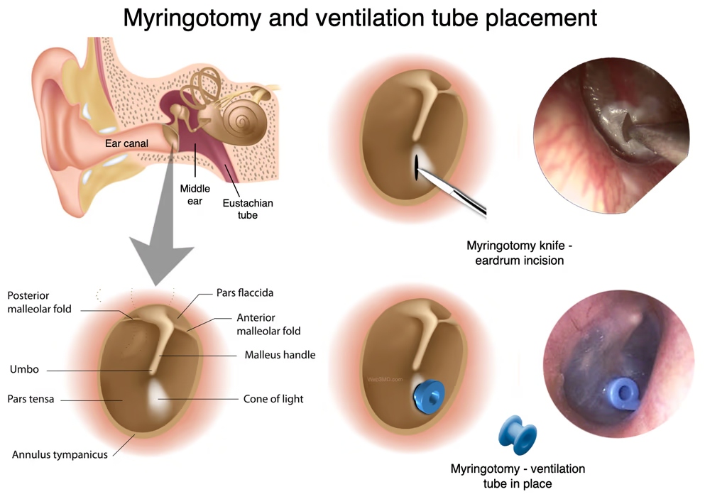
– Otitis media with effusion: it often improves with active observation, allergy management, and nasal hygiene; when it persists with an impact on hearing, ventilation tubes may restore conductive hearing, and adenoidectomy may be considered in specific situations. In parent-friendly language, understand why an ear tube is sometimes needed.
– Conductive hearing loss: earwax removal, treatment of ear infections, and bone-conduction devices can offer meaningful functional gains.
– Sensorineural hearing loss: modern hearing aids and remote-microphone systems improve speech understanding at home and at school; in profound bilateral loss, a cochlear implant, after careful evaluation, can open valuable opportunities for language.
– Congenital cytomegalovirus: in selected scenarios, antivirals started early may reduce the progression of hearing loss, always under specialized care.
– Speech therapy and school: family-centered therapy, auditory-verbal stimulation, and classroom adjustments help consolidate outcomes and independence.
– Ongoing follow-up: children with risk factors or fluctuating hearing loss need periodic reassessments, device adjustments, and consistent communication strategies.
Prevention and healthy habits
– Keeping vaccinations up to date reduces infections associated with hearing loss.
– Managing rhinitis and allergies lowers the number of episodes of otitis media with effusion.
– Responsible use of headphones, regular listening breaks, and comfortable volumes help protect the cochlea; at concerts and motorsports, ear protection is recommended.
– Environments with good acoustics at home and at school support speech understanding and learning.
When to seek an evaluation
Delayed speech, recurrent ear infections, difficulty in noisy environments, or a failed result at any stage of screening are reasons to consult an otolaryngologist (ENT) and a speech-language therapist. The earlier care begins, the greater the gains in language, academic performance, and social well-being.
Excellent care in São Paulo and Alphaville
Dr. José Eduardo Marcondes offers comprehensive, compassionate care for hearing loss in children. With more than two decades of experience and practice at leading institutions, each child receives a tailored plan, with clear communication, close follow-up, and treatment goals aligned with the family’s routine.
Next step
Whenever there is any concern, scheduling a specialized evaluation is the safest way to protect a child’s development. An attentive consultation, with age-appropriate tests, makes it possible to act with precision and confidence, so that the child can grow up hearing, speaking, and learning to the fullest.
Dr. José Eduardo Marcondes, physician, otolaryngologist (ENT). CRM-SP 107.711 (Brazilian medical license) | RQE 43.840 (specialist registration). This content is informational, does not replace a medical consultation, and does not supersede the plan of care defined by your own physician.
Trained and completed his residency at Escola Paulista de Medicina (UNIFESP), with more than two decades of experience. Focused on the treatment of snoring and sleep apnea, nasal obstruction, chronic sinusitis, adenoids and tonsils, in adults and children. Member of ABORL-CCF and of the medical staff at Hospital Israelita Albert Einstein, Vila Nova Star and São Luiz.
How to recognize sleep apnea in children: signs parents should not ignore
As parents, you spend hours watching your children sleep, taking in every detail of that relaxed little face and their breathing. But have you ever noticed any unusual behavior during sleep? Loud snoring, pauses in breathing, restless sleep or excessive daytime sleepiness may be signs of obstructive sleep apnea, a problem more common than many people realize and one that can seriously affect a child’s development. To put it in perspective, obstructive sleep apnea affects about 1% to 5% of children and is most common between ages 2 and 8, when the tonsils and adenoids tend to be proportionally larger.
What is sleep apnea in children?
Obstructive sleep apnea is a breathing disorder that occurs during sleep, characterized by brief interruptions in breathing. During these pauses, which may last a few seconds, the child essentially stops breathing because of a blockage in the upper airway. These episodes happen repeatedly throughout the night, fragmenting sleep and preventing the child from getting the restorative rest that their development requires.
Recognizing changes in everyday behavior
Often, the first signs of apnea appear during the day, in the child’s everyday activities. As a physician, I always ask parents to observe how their children behave at school. Children with apnea frequently have trouble concentrating in class, getting easily distracted during explanations or while doing their homework.
Memory can also be affected. You may notice that your child forgets simple messages, has trouble remembering where they left their toys, or needs several repetitions to learn a song or a poem. Information processing tends to slow down, so the child may take longer to understand instructions or answer questions.
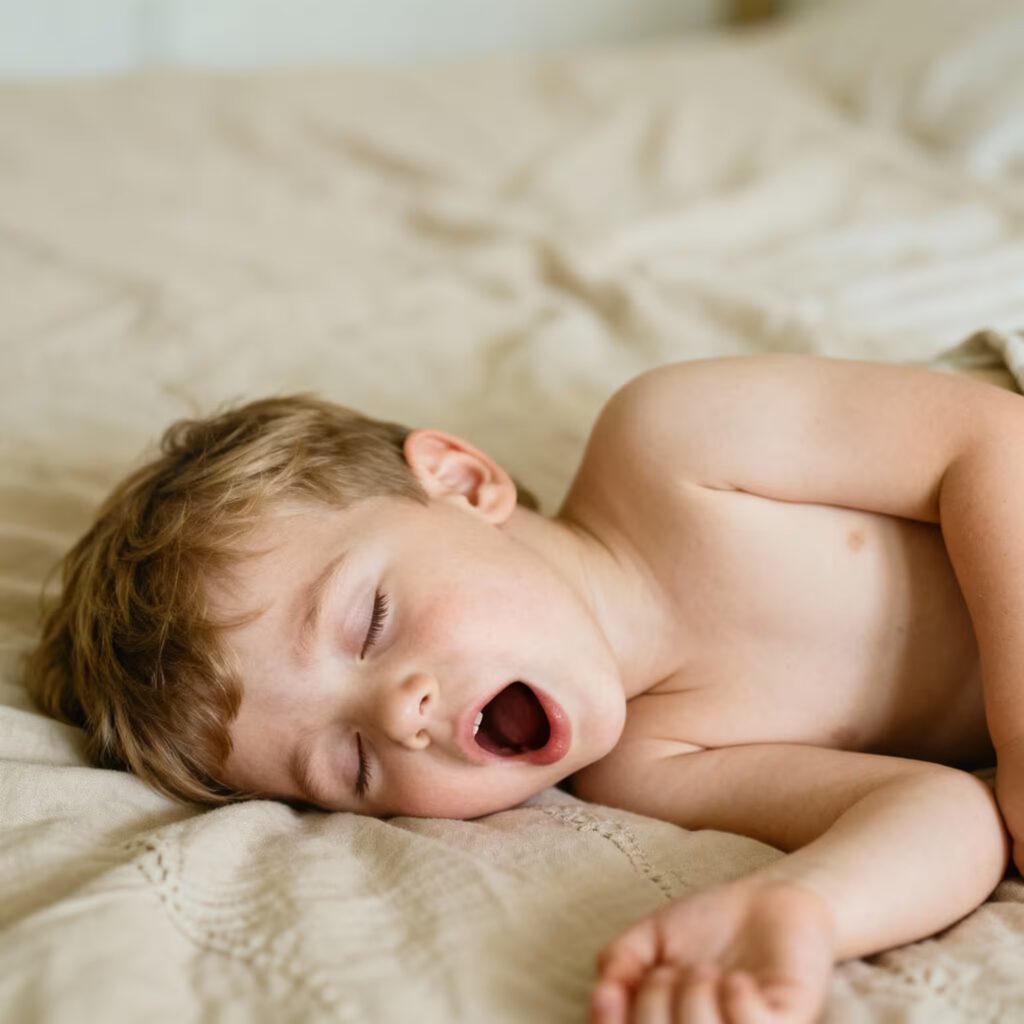
Child with daytime sleepiness lying down yawning
In terms of behavior, watch for excessive irritability, especially toward the end of the day. Children who were once calm may become aggressive, have tantrums for no apparent reason, or show signs of hyperactivity. Paradoxically, unlike adults who tend to feel sleepy, many children with apnea become agitated and restless as a way of compensating for their tiredness.
School performance is often one of the first things to catch the attention of parents and teachers. Falling grades, difficulty keeping up with the class, and problems with reading and writing can be direct consequences of fragmented sleep. Some children are even referred for an attention deficit evaluation when the real problem lies in inadequate sleep.
How fragmented sleep harms development
When sleep is constantly interrupted by apnea episodes, the child cannot reach the deeper stages of sleep that are essential for development. During these stages, the brain consolidates memories, processes information learned during the day and releases hormones that are fundamental for growth.
Chronic oxygen deprivation, caused by the breathing pauses, has a direct impact on how the brain functions. Brain cells, especially in the areas responsible for attention, memory and learning, are affected by this reduced oxygen supply. Over time, and if left untreated, this may lead to lasting changes in cognitive development.
In addition, the child’s body remains in a constant state of stress during the night, releasing hormones such as cortisol that can interfere with growth and emotional development. This is why many children with apnea show poor weight and height gain, along with mood changes.
Signs that should get your attention
As a physician with more than 20 years of experience, I always encourage parents to pay close attention to how their children behave both at night and during the day.
During sleep, watch for frequent, loud snoring, which can be intense enough to wake them at night. Notice whether there are pauses in breathing followed by gasping or choking sounds. The child may sleep in unusual positions in an attempt to ease the passage of air, have extremely restless sleep with constant changes of position, and sweat excessively during the night.
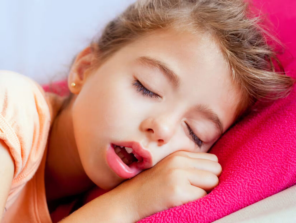
Child with sleep apnea sleeping with the mouth open
During the day, notice whether the child shows excessive sleepiness, constant tiredness even after a full night’s sleep, irritability, aggressiveness or hyperactivity, difficulty concentrating at school and a drop in school performance. Often, unlike adults who feel sleepy, children with apnea may become hyperactive and restless.
Specific signs related to breathing and eating
Mouth breathing is an important sign that should not be ignored. When the child keeps their mouth constantly open, even during the day, it may indicate chronic nasal obstruction. Persistent bad breath and dryness of the lips and around the mouth are also consequences of this oral breathing.
At mealtimes, watch for specific behaviors that point to breathing difficulty. Children with apnea frequently eat with their mouth open, making noises while chewing. They may chew very slowly, stopping repeatedly to breathe through the mouth, or swallow large pieces of food to avoid chewing for too long.
Some children prefer soft or liquid foods, avoiding textures that require prolonged chewing. During meals, they may seem breathless or tired, especially when eating foods that take more chewing effort. It is also common for them to complain of difficulty swallowing or a sensation of food being “stuck” in the throat.
Also pay attention to complaints of a blocked ear, ringing in the ears or frequent ear pain. Apnea can cause Eustachian tube dysfunction, leading to recurrent ear infections and an impact on hearing.
Why is it so important to stay alert?
Sleep apnea is not just a matter of breathing during the night. Its consequences can be serious for a child’s development, which is why I always stress the importance of early diagnosis.
Impact on growth and development: During deep sleep, the body releases hormones that are essential for growth. Children with apnea may show poor weight and height gain, along with changes in facial development and the dental arch.
Cognitive and learning difficulties: Fragmented sleep and reduced oxygen levels directly affect brain development. Studies show that children with apnea have difficulties with memory, attention, concentration and information processing. School performance may suffer, with lower grades and learning difficulties.
Behavioral and emotional changes: A lack of quality sleep can lead to significant behavioral problems. Children may show hyperactivity, aggressiveness, attention deficit, irritability and even symptoms resembling ADHD. Emotional problems such as anxiety and depression may also develop.
Cardiovascular complications: Although less common in children, apnea may lead to pulmonary hypertension and, in severe cases, heart problems. Chronic oxygen deprivation places an added burden on the cardiovascular system.
Important risk factors
Some children are more prone to developing sleep apnea. Enlarged tonsils and adenoids are the most common cause in childhood. Childhood obesity also significantly increases the risk. Children with respiratory allergies, gastroesophageal reflux, craniofacial malformations or genetic syndromes also deserve special attention. When the cause is enlarged tonsils and adenoids, surgery (adenotonsillectomy) is the first-line treatment and helps most children. Even so, it is not a guaranteed cure: some children, especially those with obesity or who are older, may keep some degree of apnea after surgery. That is why re-evaluation after treatment is so important.
The importance of early diagnosis
It often takes a long time between the first symptoms and the correct diagnosis. During this time, the child is exposed to all the complications I mentioned. That is why I always tell parents: do not ignore the signs.
Diagnosis involves a detailed clinical evaluation, specific questionnaires to screen for sleep disorders and, when necessary, polysomnography (sleep study), which is the gold-standard test. The earlier we identify and treat apnea, the better the outcomes for the child’s development.
What to expect from treatment
A large international trial (the CHAT trial, published in the New England Journal of Medicine in 2013) helped clarify what to expect from surgery in children with mild to moderate apnea. Compared with watchful waiting, adenotonsillectomy improved behavior, quality of life, symptoms, and the sleep study itself.
How it is treated
Treatment is individualized. In milder cases, measures such as a nasal corticosteroid spray and, in selected situations, a leukotriene antagonist (montelukast) may help, although the evidence is short-term. When the tonsils and adenoids are significantly enlarged, surgery is the first choice. If apnea persists after surgery, or when surgery is not indicated, CPAP (a device that keeps the airway open with air pressure) is a good option. And in a child with obesity, weight control is part of treatment, because obesity raises the risk that apnea persists.
When to seek help?
If you notice any of these signs in your children, especially frequent snoring, breathing pauses observed during sleep or significant behavioral changes, do not hesitate to seek a specialist evaluation. As I always tell parents in my office: the quality of your child’s sleep is fundamental to their future.
Keep in mind that sleep apnea in children can be treated, and the earlier the intervention, the better the outcomes for the child’s physical, cognitive and emotional development. Schedule an appointment through our WhatsApp so that, together, we can find the best way to ensure peaceful nights and healthy development for your little one.
Frequently asked questions
Is every snore a sign of apnea?
No. Occasional snoring is common. The warning sign is frequent, loud snoring together with pauses in breathing, gasping, or very restless sleep. It is that combination that warrants evaluation.
Does my child really need a sleep study (polysomnography)?
Polysomnography is the standard test to confirm apnea and measure its severity. Not every child needs it right away: the assessment starts with the clinical history and an exam of the nose and throat, and the specialist orders it when the result will change management.
Does tonsil and adenoid surgery cure apnea?
In most children with enlarged tonsils and adenoids, surgery greatly improves or resolves the problem. But it is not a guarantee: children with obesity or who are older may keep some degree of apnea, so re-evaluation after surgery is important.
My child is obese. Does that change anything?
Yes. Obesity increases the risk of apnea and the chance it persists even after surgery. In these cases, weight management is part of treatment and CPAP is sometimes needed as well.
Can apnea be mistaken for ADHD?
It can. Apnea causes inattention, restlessness, and hyperactivity that resemble ADHD, and children are sometimes referred as if they had the disorder. Treating the apnea often improves these symptoms, but the two conditions can also coexist. That is why it is worth investigating sleep before making the diagnosis.
References
Marcus CL, et al.; CHAT. A randomized trial of adenotonsillectomy for childhood sleep apnea. N Engl J Med. 2013;368(25):2366-2376.
Marcus CL, et al.; AAP. Diagnosis and Management of Childhood Obstructive Sleep Apnea Syndrome. Pediatrics. 2012;130(3):576-584.
Lumeng JC, Chervin RD. Epidemiology of pediatric obstructive sleep apnea. Proc Am Thorac Soc. 2008;5(2):242-252.
Bhattacharjee R, et al. Adenotonsillectomy outcomes in treatment of obstructive sleep apnea in children: a multicenter retrospective study. Am J Respir Crit Care Med. 2010;182(5):676-683.
Kheirandish-Gozal L, Gozal D. Intranasal budesonide treatment for children with mild obstructive sleep apnea syndrome. Pediatrics. 2008;122(1):e149-e155.
Goldbart AD, Greenberg-Dotan S, Tal A. Montelukast for children with obstructive sleep apnea. Pediatrics. 2012;130(3):e575-e580.
Dr. José Eduardo Marcondes, physician, otolaryngologist (ENT). CRM-SP 107.711 (Brazilian medical license) | RQE 43.840 (specialist registration). This content is informational, does not replace a medical consultation, and does not supersede the plan of care defined by your own physician.
Trained and completed his residency at Escola Paulista de Medicina (UNIFESP), with more than two decades of experience. Focused on the treatment of snoring and sleep apnea, nasal obstruction, chronic sinusitis, adenoids and tonsils, in adults and children. Member of ABORL-CCF and of the medical staff at Hospital Israelita Albert Einstein, Vila Nova Star and São Luiz.
What should parents watch for? When is surgery needed?
I want to talk to parents about adenotonsillectomy in children. I know this topic can raise many questions, so I will explain in a simple and clear way everything that is important for you to understand what is happening with your child, what to watch for, when it may be time to consider surgery, and how it is performed.
First of all: what are the adenoids?
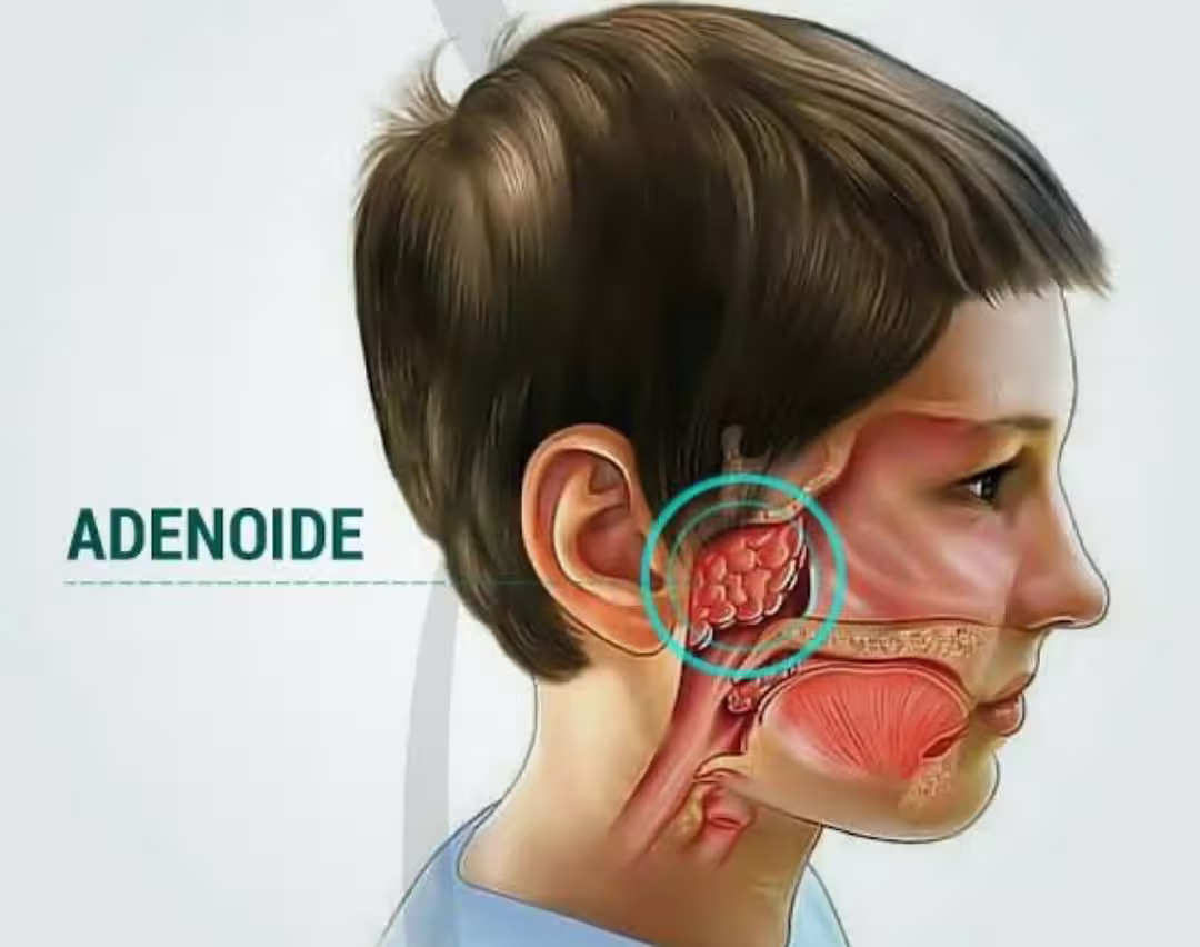
This is one of the most common questions parents ask. What are the adenoids, sometimes called “spongy tissue”? The adenoids are a type of tonsil located at the back of the nose. Every child has this structure, but it can cause significant problems when it becomes too large.
What aspects should you pay attention to?
One of the most important things to notice is whether your child frequently breathes through the mouth, even when not having a cold. Blocked nasal breathing causes the child to sleep with the mouth open and often to snore.
Also keep an eye on your child’s mood: excessive tiredness, irritability with no apparent cause, and lack of energy may be linked to poor oxygenation during sleep.
If you notice a more “muffled,” nasal-sounding voice, this may be a sign of nasal obstruction. It is also worth noting any reports of pauses in breathing while the child sleeps.
Another sign is children who eat very quickly or take a long time to swallow their food.
Symptoms and signs that may indicate enlarged adenoids
When the adenoids grow too much, certain signs tend to appear that do not go unnoticed.
The child complains of a stuffy nose without having a cold, has persistent bad breath, and dryness around the mouth.
Children often complain of a “blocked ear” or ringing, because enlarged adenoids may impair proper drainage of the middle ear, which can favor infections. Recurrent ear infections are common and may affect hearing.
During sleep, in addition to snoring, restless sleep is frequent, with the child waking several times, which can lead to drowsiness or difficulty concentrating at school.
When is surgery indicated?
I usually recommend adenotonsillectomy when treatment with nasal sprays, antibiotics, or clinical follow-up does not resolve the obstruction and infections keep recurring.
If your child snores a lot, has obstructive sleep apnea at night, or has had three or more ear infections within a few months, surgery may be the solution in selected cases.
In addition, prolonged mouth breathing may affect facial and dental development, which is why timely intervention can help prevent changes in the dental arch and tooth alignment.
Traditional surgical techniques for adenoids and tonsils in children
The classic method of removing the adenoids and tonsils in children uses instruments to cut and curette the tissue. It is a safe procedure, but it may cause somewhat more discomfort and bleeding.
Electrocautery, which uses heat to remove the tissue and control bleeding, has become popular because it reduces intraoperative bleeding. However, it can still cause moderate pain in the first few days after surgery.
Advanced technologies: microdebrider and radiofrequency
To offer children greater comfort and safety, I use two state-of-the-art technologies.
The microdebrider for adenoidectomy is used to perform the surgery with video guidance, allowing only the excess tissue to be removed, healthy structures to be preserved, and bleeding to be reduced.
In tonsillectomy, radiofrequency removes the tissue with controlled heat, which tends to result in less postoperative pain and faster recovery. Recovery is generally far more comfortable with this technology.
These technologies may allow earlier discharge, a quicker return to school activities, and much greater peace of mind for the whole family.
If your child shows these characteristics, schedule a consultation through our WhatsApp to better understand the best options to improve your little one’s quality of life.
Dr. José Eduardo Marcondes, physician, otolaryngologist (ENT). CRM-SP 107.711 (Brazilian medical license) | RQE 43.840 (specialist registration). This content is informational, does not replace a medical consultation, and does not supersede the plan of care defined by your own physician.
Trained and completed his residency at Escola Paulista de Medicina (UNIFESP), with more than two decades of experience. Focused on the treatment of snoring and sleep apnea, nasal obstruction, chronic sinusitis, adenoids and tonsils, in adults and children. Member of ABORL-CCF and of the medical staff at Hospital Israelita Albert Einstein, Vila Nova Star and São Luiz.
A complete guide to help you understand snoring and sleep apnea, whether surgery can help, and how it works.
Starting out: what causes snoring? What is sleep apnea? How does it happen?
Sleep apnea is a pause in breathing that occurs during the night. Snoring is a noise produced by the vibration of the airway structures as air passes through. But what does one have to do with the other? Why does this happen?
Let’s go back a little so this becomes clearer. Let’s quickly understand how our breathing works, because from there it becomes very easy to understand what snoring and sleep apnea are.
When we breathe in, in order for air to enter the body, the muscles of the lungs, especially the diaphragm, expand the rib cage, increasing its volume (you can see your chest expanding as you inhale). This expansion increases the volume of the rib cage and, as a result, lowers the pressure inside the lungs. This negative pressure spreads throughout the airway (trachea, larynx, pharynx, oral cavity, and nose), and air flows into the lungs due to the pressure difference. Pay attention to this concept, because it will be essential to understanding what happens in your throat that makes you snore while you sleep.
When we sleep, the muscles of the body relax, and consequently the muscles of the pharynx and tongue also become more lax. As we draw in air during the night, the negative pressure created by the lungs travels throughout the airway and meets the relaxed muscles. Depending on the intensity of the airflow and the degree of muscle relaxation, these muscles vibrate and act like a kind of musical instrument, producing a sound, the well-known snore. So snoring is nothing more than the noise produced by the vibration of the airway muscles. Now, depending on the intensity of the negative pressure created, the airway does not merely vibrate, it can close, blocking the passage of air and causing what we call apnea. So you can see how sleep apnea and snoring are closely linked. They are different points on the spectrum of the same process.
If apnea and snoring are so closely linked, what is the difference between them?
The main difference lies in the impact on health that each of these problems can cause. Sleep apnea restricts the passage of air, either fully or partially, and this can lead to a range of harmful effects on the body, such as changes in blood pressure, the endocrine system, and immunity, as well as diabetes, an increased risk of heart attacks and strokes, impotence, among others. It is worth noting that treating apnea has been shown to improve symptoms, sleep, and quality of life (SAVE trial, 2016). If you want to understand exactly what happens, read the following article:
Isolated snoring, known as primary snoring, may not seem like a major problem for most people, since its main complication is social. You cannot sleep next to your partner, you feel embarrassed traveling with friends, you cannot sleep at all on a plane.
But the issue runs deeper. Snoring is not the same as having apnea. Snoring is a warning sign that deserves evaluation, not a diagnosis in itself. For this reason, people who snore habitually should be investigated, especially when associated signs are present. Compared with the general population, snorers are about 1.5 to 3 times more likely to have sleep apnea, depending on the diagnostic criteria and the sample.
Sleep apnea is more common than it seems. In the EPISONO study, conducted in the city of São Paulo, about one third of adults (32.8%) had some degree of sleep apnea. Severity is measured by the AHI, the apnea-hypopnea index, which is the number of breathing pauses per hour of sleep: up to 5 is considered normal, 5 to 15 is mild, 15 to 30 is moderate, and above 30 is severe.
Certain features make this evaluation even more important. Here is what they are:
Frequency: if you snore more than 3 to 4 times a week
Associated symptoms: morning headaches, difficulty concentrating and memory changes, daytime sleepiness, irritability, loss of libido
Presence of other conditions: high blood pressure, diabetes, cardiovascular disease, erectile dysfunction
Other factors: low testosterone, obesity, sleep medications
The evaluation for sleep apnea is always carried out through polysomnography (a sleep study), and also through certain additional tests that may be important depending on the case and the planned course of treatment, such as a CT scan of the paranasal sinuses, a CT scan of the neck, flexible nasolaryngoscopy, and drug-induced sleep endoscopy, among others.
What is the treatment for sleep apnea?
The treatment of sleep apnea must be assessed on a case-by-case basis, but the goal of each option is always the same: to keep the airway open and ensure that air can pass through it.
Each form of treatment uses different mechanisms to achieve this, and the main options that may be used, alone or in combination, are:
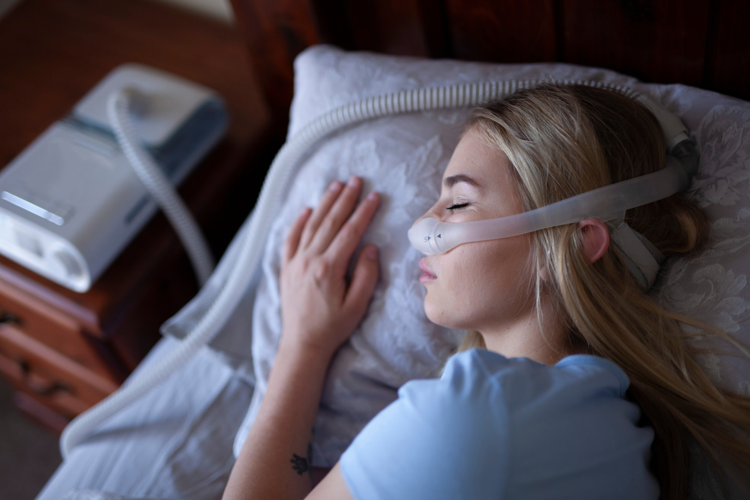
CPAP: this device creates positive air pressure, reversing the mechanics of breathing. In other words, instead of the lungs creating negative pressure to “pull” air in, the CPAP creates external positive pressure and “pushes” air into the body.
Woman with sleep apnea sleeping with a CPAP
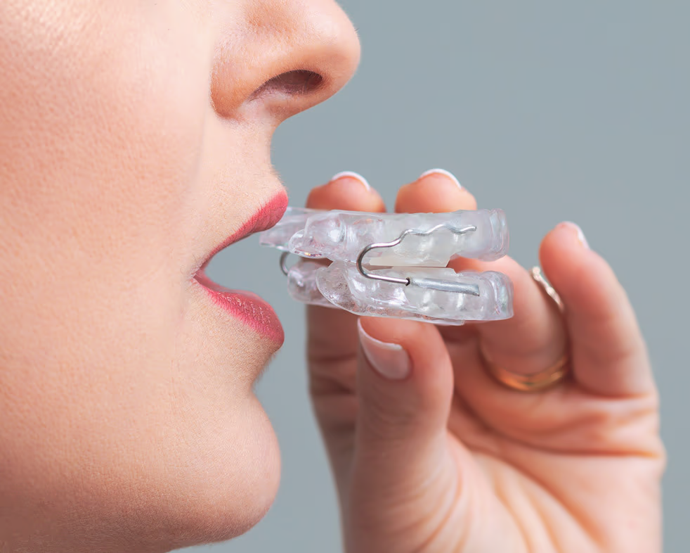
Oral appliance: this device pulls the lower jaw forward and repositions the tongue and pharynx, keeping the airway open.
Oral appliance opening the airway of a person with sleep apnea
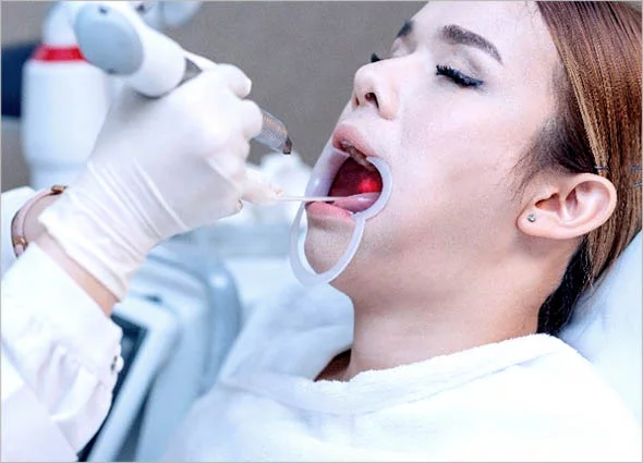
Speech therapy: through specific exercises for the tongue, palate, pharynx, neck, and chest, it is possible to strengthen the muscles and help maintain a clear airway that favors the passage of air.
Speech therapist assessing the palate of a patient with sleep apnea
Fotona laser: stimulates collagen production and the tightening of throat tissues to open the airway
Fotona laser being used to treat sleep apnea
Surgery: by removing and repositioning various structures, it provides a wider airway that withstands the pressure of breathing, keeping air flowing through it
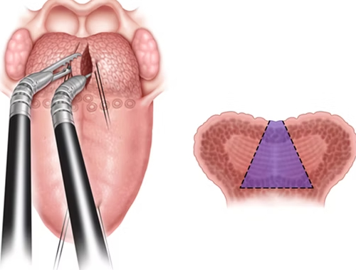
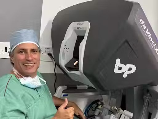
Sleep apnea surgery performed with the assistance of the Da Vinci robotSleep apnea surgery using barbed reposition pharyngoplasty
Sleep apnea surgery: How is it performed? What are the results?
A key concept: it is not just one surgery, it is several
Surgery is an important option for patients who have not adapted to CPAP or other treatments. CPAP is the first-line treatment and is very effective when well tolerated, but between 30% and 50% of people do not adapt to it or abandon the device over time, and that is where surgery has a role. What many people do not realize is that this is not a single surgery, but rather a set of procedures performed at the same time, each directed at a specific point of the upper airway.
The Concept of Multiple Procedures
The main goal of surgical treatment for sleep apnea is to keep the airway open during sleep. Because obstructive events can occur at different levels of the upper airway (nose, palate, lateral pharyngeal walls, and base of tongue), the most effective surgical approach involves treating multiple areas at the same time. This combined strategy has been shown to produce better results compared with treating a single site of obstruction.
Nasal Level: Clearing the Upper Airway
Septoplasty: Correction of a Deviated Septum
Correcting a deviated nasal septum is often the first step in multilevel surgery. A deviated septum can increase resistance to airflow through the nose and encourage mouth breathing during sleep, which worsens snoring. It is worth being clear about the role of the nose: nasal obstruction tends to worsen snoring and make CPAP use more difficult, but correcting the nose alone rarely cures apnea. It improves breathing, snoring, and often CPAP tolerance, which is why it is usually the first step of a multilevel surgery.
During septoplasty, we make a small incision inside the nose, where the mucosa is lifted away from the cartilage and bone of the septum. The deviated portions are then removed or repositioned to center the nasal septum. This procedure significantly improves nasal airflow and reduces the tendency toward mouth breathing during sleep.
Turbinoplasty: Reduction of the Nasal Turbinates
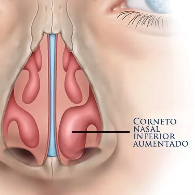
Surgery on the nasal turbinates is often performed together with septoplasty. Enlarged turbinates can cause chronic nasal obstruction, forcing the patient to breathe through the mouth during sleep, which destabilizes the upper airway. Turbinoplasty aims to reduce the volume of the inferior turbinates while preserving their physiological function of filtering and humidifying the air.
Ethmoidectomy and Maxillary Sinus Surgery
When necessary, we also perform ethmoidectomy and maxillary sinus surgery. Ethmoidectomy involves removing inflamed tissue, polyps, or blockages in the ethmoid sinuses that prevent proper ventilation. This procedure, performed using an endoscopic technique, ensures precision and minimizes the surgical impact. Maxillary sinus surgery complements the treatment when the maxillary sinuses are affected, ensuring proper drainage and reducing recurrent infections that may contribute to nasal obstruction.
In addition, combining this procedure helps create a second airflow pathway that can reduce airway resistance, contributing to the success of the surgery as a whole.
Palatal Level: Enlarging the Retropalatal Space
Barbed Reposition Pharyngoplasty to Open the Palate
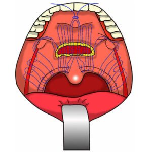
Barbed reposition pharyngoplasty is a modern, minimally invasive technique for treating the soft palate. This technique uses special sutures with small bidirectional barbs that reinforce and stabilize the throat tissues, improving the tension of the upper airway.
Each barb of the suture acts as a knot anchored in the tissue, providing greater airway stability with shorter operating time and better healing. This approach is particularly effective for patients whose snoring is caused by excessive relaxation of the palatal tissues, offering faster recovery and good results.
Lateral Wall Level: Functional Expansion with the Da Vinci Robot
Functional Expansion Pharyngoplasty with the Da Vinci Robot
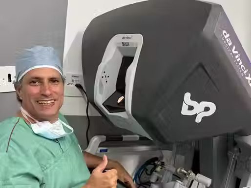
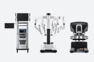
A major evolution of this technique is Functional Expansion Pharyngoplasty performed with the Da Vinci robotic system. This procedure combines the principles of traditional expansion pharyngoplasty with the precision and advantages of transoral robotic surgery.
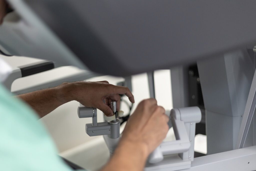
The robotic functional expansion technique uses magnified 3D visualization and articulated instruments with 360-degree movement to reposition the palatopharyngeus muscle. During the procedure, the palatopharyngeus muscle is isolated from the mucosa and from the superior pharyngeal constrictor muscle, and is then divided inferiorly to form a muscle flap with a superior and medial pedicle.
The free end of the palatopharyngeus muscle is then rotated upward and fixed at the junction between the soft and hard palate, creating tension in the lateral pharyngeal wall. The robot allows this fixation to be carried out with millimeter precision, ensuring optimal positioning of the muscle and reducing the risk of complications.
This robotic approach to functional expansion offers advantages over the traditional technique. The three-dimensional visualization allows precise identification of the anatomical structures, while the articulated instruments make it possible to suture at angles that would be impossible with conventional techniques. The result is a more consistent enlargement of the pharyngeal space, with good results in appropriately selected patients.
Base of Tongue Level: Robotic Surgery
Robotic Lingual Tonsillectomy
Transoral robotic surgery (TORS) using the Da Vinci system represents the most advanced development in the treatment of base of tongue obstruction.
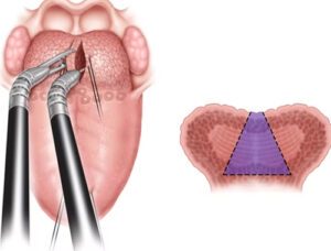
Robotic lingual tonsillectomy aims to remove enlarged tissue at the base of the tongue, including the lingual tonsils. The procedure may remove up to 20 grams of tissue as needed, based on the patient’s anatomy and the degree of collapse during sleep.
Partial Glossectomy
Midline partial glossectomy complements robotic treatment when there is significant macroglossia. This procedure involves the physical removal of a portion of the tongue in the central area, between the major blood vessels and nerves. The technique is guided by advanced imaging methods to ensure maximum safety and to preserve the essential functions of the tongue.
Advantages of the Combined Approach
Performing these multiple procedures at the same time offers several advantages. First, it makes it possible to treat all levels of obstruction in a single surgical session, reducing the need for subsequent surgeries. In addition, the multilevel approach tends to achieve higher success rates than treating a single site.
Robotic surgery offers specific benefits, including less intraoperative bleeding, faster recovery, and functional preservation with a lower risk of changes in swallowing and speech. The high-definition 3D visualization and the precision of the articulated instruments help prevent bleeding and allow removal of the excess tissue.
Recovery and Results
Recovery varies according to the extent of the procedures performed.
Multilevel surgery improves apnea in most well-selected cases. In meta-analyses, the success rate (a reduction in the AHI of at least half, with a final AHI below 20) is around 60% to 66%, with an average reduction of about 25 events per hour. Barbed reposition pharyngoplasty and robotic base-of-tongue surgery reduce the AHI by around 24 events per hour in the published series. It is worth being transparent: results depend heavily on patient selection and on the site of obstruction, and surgery usually improves symptoms and quality of life considerably, without necessarily eliminating apnea entirely.
Combined surgery for the treatment of sleep apnea therefore represents a comprehensive solution that treats the points of upper airway obstruction at the same time. This multilevel approach offers patients an effective alternative to CPAP, with lasting results and a significant improvement in quality of life.
Frequently asked questions
Does everyone who snores have sleep apnea?
No. Snoring is a warning sign that deserves evaluation, especially if it is frequent or comes with daytime sleepiness, morning headaches, or high blood pressure. The chance of apnea is about 1.5 to 3 times higher in people who snore than in the general population, but snoring by itself is not a diagnosis of apnea.
How is the severity of apnea measured?
By the AHI (apnea-hypopnea index), which counts the pauses in breathing per hour of sleep, measured during polysomnography (the sleep study): up to 5 is normal, 5 to 15 is mild, 15 to 30 is moderate, and above 30 is severe.
Does surgery cure sleep apnea?
In most well-selected cases, surgery greatly improves apnea and quality of life, but it does not always eliminate the problem. Success depends on the site of obstruction and on each patient’s characteristics, which is why a detailed evaluation before surgery is so important.
Do I need to have all of these surgeries?
No. Apnea can cause obstruction at different points (nose, palate, pharyngeal walls, base of the tongue), and surgery is individualized: only what is needed is treated, often in a single surgical session.
Does surgery replace CPAP?
CPAP is the first-line treatment and is very effective when well tolerated. Surgery is the main alternative for those who do not adapt to the device or abandon it, and it is sometimes used in combination with other measures.
References
Senaratna CV, et al. Prevalence of obstructive sleep apnea in the general population: a systematic review. Sleep Medicine Reviews. 2017;34:70-81.
Wali SO, Abalkhail B, Krayem A. Prevalence and risk factors of obstructive sleep apnea syndrome in a Saudi Arabian population. Annals of Thoracic Medicine. 2017;12(2):88-94.
Tufik S, et al. Obstructive Sleep Apnea Syndrome in the São Paulo Epidemiologic Sleep Study (EPISONO). Sleep Medicine. 2010.
McEvoy RD, et al. CPAP for Prevention of Cardiovascular Events in Obstructive Sleep Apnea (SAVE trial). New England Journal of Medicine. 2016.
Dr. José Eduardo Marcondes, physician, otolaryngologist (ENT). CRM-SP 107.711 (Brazilian medical license) | RQE 43.840 (specialist registration). This content is informational, does not replace a medical consultation, and does not supersede the plan of care defined by your own physician.
Trained and completed his residency at Escola Paulista de Medicina (UNIFESP), with more than two decades of experience. Focused on the treatment of snoring and sleep apnea, nasal obstruction, chronic sinusitis, adenoids and tonsils, in adults and children. Member of ABORL-CCF and of the medical staff at Hospital Israelita Albert Einstein, Vila Nova Star and São Luiz.
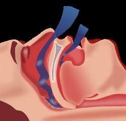
During obstructive sleep apnea, the upper airway collapses in the region of the pharynx. When you sleep, the throat muscles relax naturally, but in people with apnea this relaxation is excessive, causing the airway to close partially or completely.
Imagine a straw that collapses as you try to breathe through it. Your body keeps working to breathe, creating negative pressure in the chest, but air cannot get through. During each episode, which lasts at least 10 seconds (and may extend beyond a minute), the body enters a state of asphyxia.
What Happens During Each Breathing Pause
When breathing stops, two critical things happen in your body:
Hypoxia: The amount of oxygen in the blood drops sharply. It is as if every cell in your body began to “starve” for oxygen.
Hypercapnia: Carbon dioxide (CO₂) builds up in the blood, creating a toxic environment. It is as if you were breathing stale air.
These events lead special sensors in your body (called chemoreceptors) to detect the danger and send urgent signals to the brain: “You need to breathe now!”. The brain then triggers a micro-arousal. You do not wake up fully, but you come out of deep sleep just enough for the throat muscles to contract and reopen the airway.
The Cascade Effect on the Cardiovascular System
Each apnea episode sets off a storm in your cardiovascular system:
The Sympathetic Nervous System on Maximum Alert
The sympathetic nervous system, responsible for “fight or flight” reactions, is intensely activated. It is a response much like being mugged or attacked by an animal. The difference is that this happens all night long, as if your body believed it were in constant danger. This causes:
A massive release of adrenaline and other stress hormones
Vasoconstriction: the blood vessels contract sharply
Rapid spikes in blood pressure: with each pause, the pressure spikes for a few seconds, which in severe cases may exceed 180 to 200 mmHg. This is not continuous high blood pressure, but this oscillation, repeated hundreds of times a night, overloads the heart and vessels over time
Apnea, the thief stealing a person’s health
The Heart Under Pressure
With each episode, your heart undergoes tremendous stress:
A sudden rise in heart rate
A higher risk of arrhythmias (irregular heartbeats)
Left ventricular hypertrophy (the heart muscle becomes “swollen” from working so hard)
An increased risk of heart attack and heart failure
How Sleep Apnea Affects Specific Body Systems
Respiratory System
Pulmonary hypertension: pressure in the lungs rises due to the lack of oxygen
Changes in the brain’s respiratory center
A gradual reduction in lung capacity over time
Endocrine and Metabolic System
Apnea can cause hormonal chaos:
Insulin Resistance: Hypoxia and stress may keep cells from responding properly to insulin, raising the risk of type 2 diabetes. It is as if the cells’ “locks” (insulin receptors) became “rusty”.
Disrupted Appetite Hormones:
Ghrelin (the hunger hormone): rises sharply
Leptin (the satiety hormone): decreases
Elevated Cortisol: The stress hormone stays high, contributing to weight gain and metabolic changes.
Nervous System
Impaired Cognitive Function:
Memory loss and difficulty concentrating
Slowed reaction time (similar to the effect of alcohol)
A higher risk of traffic accidents, about 2 to 3 times higher, especially in severe cases with marked sleepiness (CPAP treatment substantially reduces this risk)
Mood changes, depression, and anxiety
Stroke Risk: apnea nearly doubles the risk of stroke, independently of factors such as blood pressure, weight, and diabetes (study published in the New England Journal of Medicine, 2005), through the combination of hypertension, changes in cerebral blood flow, and a greater tendency to form clots.
Reproductive System
Apnea can deeply affect the sex hormones and reproductive function in both men and women:
Effects on the Male Reproductive System:
Lower Testosterone: Intermittent hypoxia and sleep fragmentation can inhibit testosterone production by suppressing the hypothalamic-pituitary-gonadal axis. Obstructive apnea is associated with lower testosterone levels, especially in more severe cases and in men with obesity. Much of this relationship, however, is explained by excess weight itself, and treating apnea alone does not always normalize the hormone.
Erectile Dysfunction: The combination of low testosterone, vascular problems, oxidative stress, and altered penile blood flow means that men with apnea may face a significantly higher risk of impotence. Apnea is considered an independent risk factor for erection problems.
Reduced Fertility: Sperm quality may be impaired by hypoxia and oxidative stress, resulting in a lower sperm count, reduced sperm motility, and increased sperm DNA fragmentation. This can make conception more difficult and raise the risk of miscarriage.
Effects on the Female Reproductive System:
Menstrual Irregularities: Apnea may cause irregular menstrual cycles due to hormonal changes, making it harder to identify the fertile window and reducing the chances of conception.
Reduced Fertility: Women with apnea may have difficulty ovulating properly due to an imbalance of the hormones FSH (follicle-stimulating) and LH (luteinizing), both essential for healthy ovulation.
Menopause-Related Problems: Apnea becomes more common after menopause due to the decline in estrogen and progesterone levels, which naturally help maintain the muscle tone of the airway. Hormone deficiency combined with apnea can create a vicious cycle that worsens menopausal symptoms.
Pregnancy Complications: Pregnant women with apnea have a higher risk of gestational hypertension, gestational diabetes, and complications during delivery.
Immune System
Sleep apnea can cause profound dysfunction of the immune system, creating a state of chronic inflammation in the body:
Persistent Inflammatory State: Intermittent hypoxia and sleep fragmentation cause the body to continuously produce pro-inflammatory cytokines such as IL-1, IL-6, and TNF-α. It is as if the body were always “fighting an infection” that does not exist.
Suppressed Immunity: The rise in cortisol and oxidative stress lead to immunosuppression, reducing the body’s ability to fight real infections. This can result in greater susceptibility to colds, flu, pneumonia, and other infectious diseases.
Imbalance of Defense Cells: There is a reduction in CD3+, CD4+, and CD8+ cells (lymphocytes important for defense) and a shift in the balance between cellular (Th1) and humoral (Th2) immune responses, favoring a pattern similar to that seen in autoimmune diseases.
Reduced NK Cell Activity: Natural Killer cells, which help defend the body, may show reduced activity. Laboratory research suggests this could weaken defenses against tumors, but it is important to be clear that this is a hypothesis under investigation: it is not proven that apnea causes cancer in people.
Oxidative Stress: The Assault of Free Radicals
One of the most damaging effects of apnea is the creation of free radicals. With each cycle of oxygen deprivation followed by reoxygenation, it is as if your cells were put through “extreme exercise,” generating toxic substances that damage:
Blood vessels (accelerating the process of atherosclerosis)
Heart cells
Neurons in the brain
Pancreatic cells (worsening diabetes)
The Vicious Cycle
Apnea creates a vicious cycle that is hard to break:
1. Apnea → Weight gain (due to altered hormones)
2. Weight gain → Worsening apnea (more tissue in the throat)
3. Worse apnea → More metabolic problems
4. More problems → More weight gain
Impact on Overall Health
Cardiovascular: about half of people with apnea also have high blood pressure, and apnea is a recognized cause of hard-to-control hypertension. Long-term studies show that severe apnea nearly triples the risk of developing hypertension. The risk of heart attack, arrhythmias, and heart failure also increases.
Metabolic: The prevalence of type 2 diabetes is much higher in patients with apnea, regardless of weight.
Neurological: A higher risk of dementia, memory loss, and accidents due to drowsiness.
Immunological: Greater susceptibility to infections. The relationship with some types of cancer is a hypothesis still under study, not a certainty.
Reproductive: Reduced fertility, sexual dysfunction, menstrual irregularities, and hormonal disturbances that affect sexual and reproductive quality of life.
Quality of Life: Excessive sleepiness, depression, relationship difficulties, and a significant drop in productivity.
The Good News
The most important point is that treating apnea may reverse many of these problems. When breathing is normalized during sleep (with CPAP, oral appliances, or surgery), the following are often observed:
A reduction in blood pressure
Improved diabetes control
A reduction in oxidative stress
Improved cognitive function and mood
Improvement in snoring, daytime sleepiness, and quality of life
Treatment reliably improves symptoms, sleep, mood and quality of life, and helps control blood pressure. Treating is well worth it for symptoms, blood pressure and quality of life.
This shows how an apparently “simple” sleep disorder can have profound consequences for the entire body, but also how appropriate treatment can transform a person’s health and quality of life. See how snoring and sleep apnea surgery works.
Frequently Asked Questions
Does sleep apnea cause cancer?
It is not proven. Laboratory research suggests that intermittent lack of oxygen may weaken part of the body’s defenses (the NK defense cells), but this is still a hypothesis under study, not a proven cause-and-effect relationship in people.
Does treating apnea prevent heart attack and stroke?
Treatment reliably improves snoring, sleepiness, mood, quality of life, and helps control blood pressure. However, the prevention of heart attack and stroke was not demonstrated by the largest randomized trial (SAVE, 2016). Even so, treating is very important for symptoms, blood pressure, and quality of life.
Does apnea raise blood pressure?
Yes. With each pause there is a rapid pressure spike, and over time apnea is a recognized cause of high blood pressure, especially the hard-to-control kind. Severe apnea nearly triples the risk of hypertension.
References
Peppard PE, Young T, Palta M, Skatrud J. Prospective study of the association between sleep-disordered breathing and hypertension. New England Journal of Medicine. 2000;342:1378-1384.
Yaggi HK, et al. Obstructive sleep apnea as a risk factor for stroke and death. New England Journal of Medicine. 2005;353:2034-2041.
Tregear S, et al. Obstructive sleep apnea and risk of motor vehicle crash: systematic review and meta-analysis. Journal of Clinical Sleep Medicine. 2009.
McEvoy RD, et al. CPAP for Prevention of Cardiovascular Events in Obstructive Sleep Apnea (SAVE trial). New England Journal of Medicine. 2016.
Su L, et al. Association between obstructive sleep apnea and male serum testosterone: a systematic review and meta-analysis. Andrology. 2022.
Dr. José Eduardo Marcondes, physician, otolaryngologist (ENT). CRM-SP 107.711 (Brazilian medical license) | RQE 43.840 (specialist registration). This content is informational, does not replace a medical consultation, and does not supersede the plan of care defined by your own physician.
Trained and completed his residency at Escola Paulista de Medicina (UNIFESP), with more than two decades of experience. Focused on the treatment of snoring and sleep apnea, nasal obstruction, chronic sinusitis, adenoids and tonsils, in adults and children. Member of ABORL-CCF and of the medical staff at Hospital Israelita Albert Einstein, Vila Nova Star and São Luiz.
If you have spent sleepless nights holding your child as they cried from an earache, and later noticed that, even without pain, they did not seem to hear well, turned the television up louder, or seemed distracted at school, you know the worry that brings you to this question. When ear infections keep coming back, or when fluid stays trapped behind the eardrum for a long time, the otolaryngologist (ENT) may suggest placing a ventilation tube, also called a tympanostomy tube. Here I have gathered the questions I hear most in the office, along with what the science shows today, so you can understand why, when, and whether this “little tube” makes sense for your child. Let me say something reassuring up front: in most cases the treatment is medical or watchful waiting, and not every child who gets ear infections will need the tube. That feeling of a blocked ear also shows up, briefly, when flying.
First, understand what the tube solves (and what it does not)
The target of treatment is the fluid trapped in the middle ear, the space behind the eardrum. It can build up after an infection (called otitis media with effusion, or OME, when there is fluid without active infection) or come along with repeated infections. While this fluid is there, the eardrum vibrates poorly and sound arrives muffled, as if the child had a blocked ear. Hence the hearing loss, ringing in the ears (tinnitus), and sometimes trouble with balance. Recurrent ear infections are a common cause of hearing loss in children.
It helps to put this in perspective, because it is less frightening once you know the whole picture. Fluid in the ear is common and, most of the time, clears on its own: about 9 in 10 children have at least one episode before age 5, half resolve within 3 months, and about 95% within a year (AAO-HNS, 2016). That is why the tube is not for every bit of fluid that appears. It comes into play when the fluid insists on staying and starts to affect hearing, or when ear infections are too frequent. What the tube does is drain that fluid and ventilate the ear, equalizing pressure. What it does not do is cure the allergy, the enlarged adenoid, or the cold behind the problem; for the cause, the treatment is different.
So, when is the tube really indicated?
Two situations concentrate the indication, and in both the ear exam matters more than the infection count alone:
Persistent fluid. Otitis media with effusion in both ears for three months or more, with documented hearing difficulty, is the best-established indication (AAO-HNS, 2022). When there are signs of impact on speech, learning, or behavior, the indication is stronger.
Recurrent ear infections, with an important caveat. Recurrent otitis media means three or more episodes in six months, or four in a year. The tube is indicated mainly when there is still fluid in the ear at an exam done between episodes; if the ear is completely dry in that period, current guidelines do not recommend the tube based on the infection count alone (AAO-HNS, 2022).
Why the caution? Because a randomized trial published in 2021 compared placing the tube with continuing medical treatment in children with recurrent ear infections and found no difference in the number of new infections over two years (1.48 vs. 1.56 episodes per child-year). The tube did have one gain: it lengthened the time until the next infection (about 4.3 vs. 2.3 months) (Hoberman, 2021). In other words, the decision is not automatic. It weighs the whole picture: hearing, the presence of fluid, the frequency of episodes, and how much all of this affects your child’s sleep, speech, and school.
Outside these situations, when the fluid is recent (less than three months) and hearing is preserved, the usual approach is to observe, because the chance of resolving on its own is high.
What is the surgery like, and is the anesthesia safe?
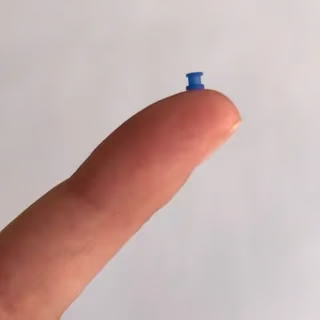
With the help of an endoscope, the doctor makes a small opening in the eardrum, suctions the fluid, and inserts the little tube, which works like a valve, letting air in and fluid out. It usually takes 10 to 15 minutes per ear, and in most cases the child goes home the same day.
The question that worries parents most is almost never about the tube, but about the anesthesia. In children we use general anesthesia, and the concern is natural. It is worth knowing that a large international randomized study followed children who received less than one hour of general anesthesia as infants and found no difference in neurological development at age 5 compared with those who did not (GAS, McCann, 2019). It is a short surgery, performed by a team used to operating on children, and this finding usually brings families peace of mind.
Will it hurt? What is recovery like?
During the surgery, thanks to the anesthesia, there is no pain. Afterward, mild discomfort or a thin discharge from the ear is common in the first few days, but nothing that usually disturbs sleep or stops play. For comfort, we recommend a simple pain reliever. If discharge appears, treatment is local, with ear drops; in most cases oral antibiotics are not needed (AAO-HNS, 2022). Within a few days the strange sensation fades, and many families notice right away that the child responds again when called. When the fluid drains, hearing improves.
Water in the ear: what has really changed
This is the point that raises the most questions, because the guidance changed. For many years, strict water protection was advised, with cotton, plugs, and caps for every bath and every pool. The most recent guidelines revised this: for most children with a tube, routine protection is not needed for ordinary bathing or for swimming in clean, treated water (AAO-HNS, 2022). Studies showed that systematic protection did not meaningfully reduce the chance of discharge.
Protection is now reserved for specific situations, such as diving deep, swimming in lake or poorly treated water, dunking the head in a soapy bath, or when the child feels discomfort from water getting in. Since every case has its particulars, your ENT will advise what applies to your child. The practical good news is that, most of the time, the bath and pool routine stays almost normal.
How long the tube stays and what to expect from the results
The tube usually stays in place for about 12 months on average, until the membrane heals around it. When the time comes, the body expels it naturally, without another surgery, and the tiny hole closes on its own in the great majority of cases. That is why follow-up visits matter: they confirm that it came out properly and that the eardrum is healthy.
As for the results, hearing usually improves quickly, and reviews of the evidence show a clear gain in the short and medium term (MacKeith, 2023).
In some cases, when improvement is incomplete, a new tube placement is needed. The main risk factors for this are early tube extrusion, craniofacial changes, younger age, and some clinical conditions such as recurrent acute otitis media (Goel, 2021). One factor that reduces the chance of another surgery is adenoidectomy performed together with tube placement, especially in children aged 4 and older (Qian, 2025); in a meta-analysis, concurrent adenoidectomy roughly halved the chance of needing new tubes (Goel, 2021).
Risks and sequelae, with numbers
This is a low-risk procedure, but, like any surgery, it has possible complications. The numbers help put them in scale (Kay, Nelson, and Rosenfeld, 2001):
Ear discharge (otorrhea): the most common, around 16% in the immediate postoperative period, almost always resolved with ear drops.
Eardrum scarring (tympanosclerosis): appears in about a third of cases after the tube comes out, but is usually just a mark, with no noticeable effect on hearing.
Perforation that does not close on its own: uncommon with short-term tubes (about 2%) and more frequent with long-term tubes; when it occurs, it can be corrected with a small repair.
Cholesteatoma: very rare (below 1%).
The tube can also become blocked or come out early, situations that are monitored in the office, where the doctor decides whether it needs to be replaced.
Are there alternatives to the tube?
Yes, and they are part of the conversation. When the fluid is recent and hearing is good, the best approach is usually to wait and reassess, because the chance of spontaneous resolution is high. Treating what lies behind the problem helps: controlling allergic rhinitis, managing repeated colds, and assessing the adenoid. In some children, especially from age 4 or when there are adenoid symptoms, removing the adenoid together with tube placement reduces the chance of new episodes (AAO-HNS, 2022). Antibiotics have a role in treating acute episodes but are not recommended as ongoing prevention. Which path makes sense depends on the exam, the hearing, and your child’s history.
In the end, the decision about the tube is shared between you and the doctor, looking at hearing, the frequency of episodes, and the impact on the child’s daily life. When well indicated, it restores good hearing, improves sleep and energy, and breaks a cycle of infections that interferes precisely with the stage of learning to speak and socialize. If any doubt remains, schedule a conversation so we can assess the case calmly. At the office of Dr. José Eduardo Marcondes, an otolaryngologist (ENT physician, CRM-SP 107.711 | RQE 43.840), guidance is always individualized, so you can decide with confidence and information.
Frequently asked questions
Can my child swim and bathe with the tube?
In most cases, yes, with no routine protection for ordinary bathing or for swimming in clean, treated water. Current guidelines no longer recommend plugs and caps for all children (AAO-HNS, 2022). Protection is reserved for specific situations, such as deep diving or poorly treated water, and your ENT advises what applies to your case.
Does the surgery hurt? And is general anesthesia safe?
There is no pain during the procedure, thanks to the anesthesia. It is a short surgery, a few minutes per ear. As for general anesthesia, a large randomized study showed that less than one hour of anesthesia early in life did not change children’s neurological development at age 5 (GAS, McCann, 2019).
Does the tube fall out on its own? Is another surgery needed to remove it?
In general the tube stays about 12 months and the body expels it naturally, without a new surgery. What matters is keeping the follow-up visits to confirm it came out and to check the eardrum.
Once placed, does it solve the problem for good?
It usually resolves the infections and hearing loss of that period. But some cases need a second tube later on, especially when the tube comes out early, in a younger child, or when there are craniofacial changes. This is expected and does not mean something went wrong.
Does every ear infection need a tube?
No. Most episodes of ear fluid resolve on their own within weeks to months. The tube is considered when the fluid persists and affects hearing, or when infections recur with fluid present at the exam.
Is there an alternative before operating?
Yes: observe and reassess when the situation is recent, treat allergic rhinitis, and assess the adenoid. In selected cases, removing the adenoid together with the tube helps reduce recurrences. The choice depends on individual assessment.
References
American Academy of Otolaryngology–Head and Neck Surgery Foundation. Clinical Practice Guideline: Tympanostomy Tubes in Children (Update). Otolaryngology–Head and Neck Surgery. 2022;166(1_suppl):S1-S55.
Rosenfeld RM, et al. Clinical Practice Guideline: Otitis Media with Effusion (Update). Otolaryngology–Head and Neck Surgery. 2016;154(1_suppl):S1-S41.
Hoberman A, et al. Tympanostomy Tubes or Medical Management for Recurrent Acute Otitis Media. New England Journal of Medicine. 2021;384(19):1789-1799.
MacKeith S, et al. Ventilation tubes (grommets) for otitis media with effusion (OME) in children. Cochrane Database of Systematic Reviews. 2023;(11):CD015215.
Kay DJ, Nelson M, Rosenfeld RM. Meta-analysis of tympanostomy tube sequelae. Otolaryngology–Head and Neck Surgery. 2001;124(4):374-380.
Goel AN, Omorogbe A, Hackett A, Rothschild MA, Londino AV 3rd. Risk Factors for Multiple Tympanostomy Tube Placements in Children: Systematic Review and Meta-Analysis. The Laryngoscope. 2021;131(7):E2363-E2370.
Qian ZJ, Truong MT, Alyono JC, Valdez T, Chang K. Tympanostomy Tube Insertion With and Without Adenoidectomy. JAMA Otolaryngology–Head & Neck Surgery. 2025;151(1):40-46.
McCann ME, et al. Neurodevelopmental outcome at 5 years of age after general anaesthesia or awake-regional anaesthesia in infancy (GAS). The Lancet. 2019;393(10172):664-677.
This content is informational, does not replace a medical consultation, and does not supersede the plan of care defined by your own physician.
Trained and completed his residency at Escola Paulista de Medicina (UNIFESP), with more than two decades of experience. Focused on the treatment of snoring and sleep apnea, nasal obstruction, chronic sinusitis, adenoids and tonsils, in adults and children. Member of ABORL-CCF and of the medical staff at Hospital Israelita Albert Einstein, Vila Nova Star and São Luiz.
Imagine waking up every morning feeling genuinely rested, with the energy to take on the day’s challenges and the readiness to enjoy every moment. This is not a distant reality or a privilege reserved for a few – it is the natural result of quality sleep, something most of us can achieve with the right habits.
Sleep goes far beyond simple rest. It is during these precious hours that the body renews itself: the immune system grows stronger, memory is consolidated, tissues repair themselves, and essential hormones are produced. When we sleep well, we not only recover energy but literally prepare ourselves to be the best version of ourselves the following day.
The Connection Between Table and Bed: How Diet Influences Sleep
What we put on our plate has a direct impact on the quality of our nights. Some foods are true allies of sleep, while others can turn bedtime into a frustrating struggle.
The stars of a sleep-friendly dinner include foods rich in tryptophan, an amino acid that the body converts into serotonin and, later, into melatonin – the well-known sleep hormone. Fish such as salmon and tuna, lean meats like chicken and turkey, eggs, milk and dairy products, along with nuts and seeds, are excellent options to include in your diet.
Fruits also deserve special mention. Bananas, in addition to tryptophan, provide magnesium and potassium, minerals that support muscle relaxation. Cherries are a natural source of melatonin, while kiwi has properties that may help both with falling asleep and with staying in deep sleep.
For dinner, the golden rule is simplicity and lightness. Very heavy meals that are rich in fat or heavily seasoned can cause digestive discomfort and disrupt sleep. Ideally, have your last meal at least two hours before lying down, so that digestion does not interfere with rest.
A few precautions are essential: avoid caffeine after 2 p.m., as it can remain active in the body for up to 8 hours. Chocolate, soft drinks, and even some teas contain stimulant substances. Alcohol, although it may initially cause drowsiness, fragments sleep during the night, resulting in poor-quality rest.
Movement That Soothes: The Role of Exercise in Sleep Quality
Regular physical activity is one of the most valuable investments we can make to improve our sleep. People who are physically active are nearly twice as likely to maintain high-quality sleep compared with those who are sedentary.
Aerobic exercise, such as walking, running, swimming, or cycling, is especially beneficial. It boosts blood circulation, promotes relaxation, and helps regulate the body’s circadian rhythms. Just 30 minutes of moderate activity can already make a meaningful difference in how easily you fall asleep and in the depth of your sleep.
The timing of exercise also matters. Activities done in the morning or afternoon help synchronize the body clock, while intense exercise too close to bedtime can have a stimulating effect. Ideally, finish vigorous workouts at least 3 hours before lying down.
For those who are short on time, gentler activities such as yoga, pilates, or stretching before bed can be excellent options. They promote muscular and mental relaxation, creating a natural transition between wakefulness and sleep.
Creating a Sanctuary for Rest: The Ideal Environment
Your bedroom should be an invitation to relax. Small adjustments to the environment can completely transform the quality of your sleep, without major expense.
Temperature is a crucial and often overlooked factor. The ideal environment for sleeping should be between 18°C and 22°C. Our body naturally lowers its temperature during sleep, and a room that is too hot or too cold can interrupt this natural process. Use bedding appropriate for the season and maintain good ventilation.
Darkness is essential for adequate melatonin production. Blackout curtains or sleep masks can be great allies, especially if you live in areas with a lot of external lighting. Avoid blue light from electronic devices for at least an hour before bed, as it “tricks” the brain into believing it is still daytime.
Silence also contributes to restorative sleep. If you cannot control external noise, consider using earplugs or relaxing sounds that mask the disturbing background noise.
The way the space is organized directly influences our ability to relax. A clean, tidy bedroom conveys a sense of calm, while cluttered surroundings can generate unconscious anxiety.
Routines That Transform: Building Powerful Habits
Consistency is the key to quality sleep. Our body works best with predictable routines, and that includes the times we go to bed and wake up.
Set a fixed time to go to bed and get up, even on weekends. This regularity helps fine-tune the internal body clock, so that you feel sleepy naturally at the right time and wake up feeling more refreshed.
Create a relaxation ritual before bed. This might include a warm bath, reading a book, meditation, or simple breathing techniques. What matters is that they are calm, pleasant activities that signal to the body that it is time to prepare for rest.
If you cannot fall asleep within 20 minutes, get out of bed and do a relaxing activity in another room, returning only when you feel sleepy. This keeps the brain from associating the bed with insomnia and anxiety.
Small Changes, Great Transformations
Improving sleep quality does not require radical changes to your routine. Small, consistent adjustments can produce surprising results. Start by choosing one or two suggestions that make the most sense for your current situation and put them into practice gradually.
Keep in mind that each person is unique, and it may take a few weeks for your body to adapt to new routines. Be patient with yourself throughout this process of change.
When we prioritize sleep, we are investing in our physical, mental, and emotional health. We are choosing more energy, a better mood, greater ability to concentrate, and a stronger immune system. In a world that often glorifies sleep deprivation as a sign of productivity, caring for your rest is an act of self-care and wisdom.
Well-slept nights are the foundation for extraordinary days. Why not start building that solid foundation today, for a fuller and healthier life?
Dr. José Eduardo Marcondes, physician, otolaryngologist (ENT). CRM-SP 107.711 (Brazilian medical license) | RQE 43.840 (specialist registration). This content is informational, does not replace a medical consultation, and does not supersede the plan of care defined by your own physician.
Trained and completed his residency at Escola Paulista de Medicina (UNIFESP), with more than two decades of experience. Focused on the treatment of snoring and sleep apnea, nasal obstruction, chronic sinusitis, adenoids and tonsils, in adults and children. Member of ABORL-CCF and of the medical staff at Hospital Israelita Albert Einstein, Vila Nova Star and São Luiz.
The nasal turbinate is a bony structure covered by mucosa located on the lateral wall of the nose, responsible for warming, humidifying, filtering, and directing the air we breathe. These functions are essential for efficient, comfortable breathing in everyday life.
Also known as the nasal concha or turbinate, it is part of the nasal cavity and is made up of bone covered by respiratory mucosa.
There are normally three turbinates on each side (inferior, middle, and superior) and, in some people, a fourth one may be present, called the supreme turbinate.
How do they help with breathing?
The aerodynamic shape of the nasal turbinates directs the airflow inside the nose, promoting a steady, quiet passage of air. Part of this flow is channeled toward the paranasal sinuses and the olfactory region, allowing us to perceive smells.
The “levels” of the nose
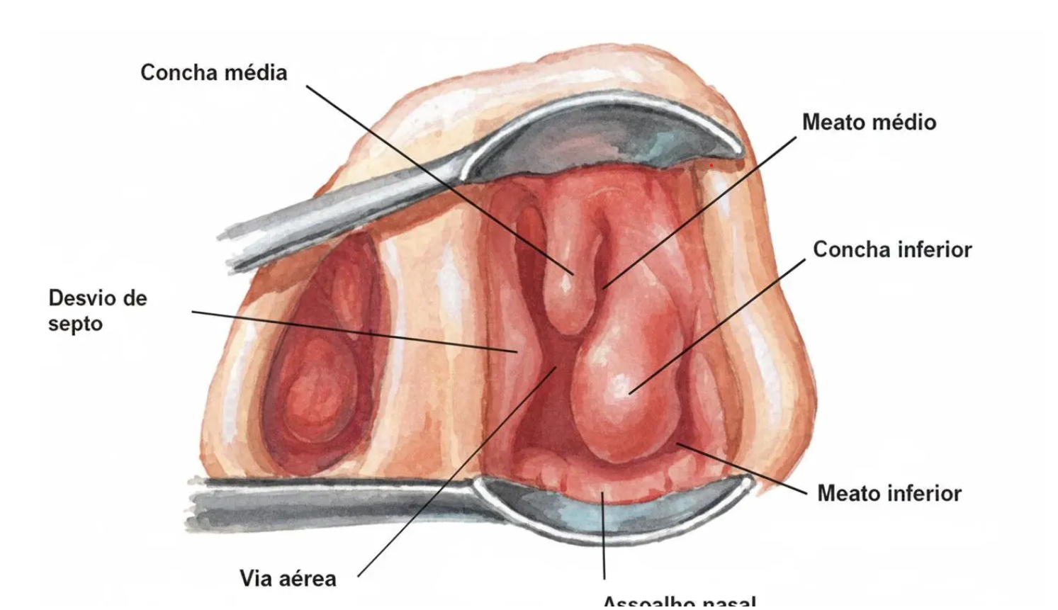
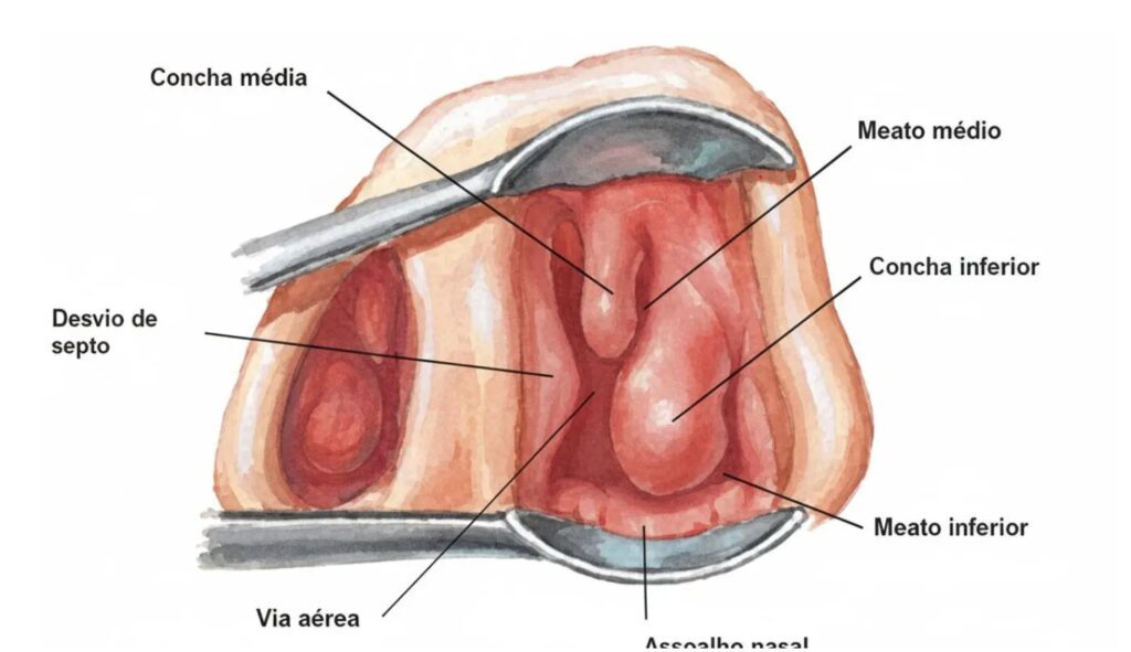
The turbinates divide the nasal cavity into meatuses, known as the “levels” of the nose: inferior, middle, and superior. In the inferior meatus, the inferior turbinate, the largest and with a rich vascular network, regulates the nasal cycle and takes part in warming and humidifying the air, and it also houses the nasolacrimal duct, through which tears drain. In other words, our tears drain into the nose, which is why the nose becomes congested when we cry.
The middle meatus contains the openings of the paranasal sinuses and tends to accumulate secretions during infections, sinusitis, and allergic processes.
The olfactory nerve, responsible for our sense of smell, is located in the superior meatus. Only a small part of the air we breathe reaches this region.
When do the nasal turbinates enlarge?
Several conditions can lead to enlargement (hypertrophy) of the turbinates and cause nasal obstruction, such as:
Rhinitis – causes chronic inflammation of the mucosa
recurrent sinusitis – infectious processes also lead to persistent inflammation of the mucosa
environmental irritants – pollutants such as sulfur, ozone, smoke particles, among others
anatomical changes – such as a deviated nasal septum, nasal polyposis, and enlarged adenoids
vascular causes – vasomotor rhinitis can also lead to enlargement of the turbinates
In adults, turbinate enlargement is often associated with other conditions, especially a deviated septum, and this combination is one of the main causes of chronic nasal congestion.
Consequences of enlarged turbinates
As we have seen, each level of the nose is responsible for a specific function, and enlargement of the nasal turbinates can interfere with each of them. The most common symptom of this enlargement is nasal obstruction, which occurs when the “lower level” is blocked. When the “second level” (known as the middle meatus) is affected, the main symptoms are related to sinus disorders, more specifically chronic sinusitis. The “third level,” the superior meatus, can lead to a reduced sense of smell.
Beyond obstruction, turbinate hypertrophy may contribute to snoring, sleep apnea, and a lower quality of breathing-related life. A feeling of being constantly congested, the need to breathe through the mouth, and worsening at night are common complaints that warrant specialized evaluation.
Medical treatment
The first line of care aims to reduce inflammation and the size of the turbinates with nasal corticosteroid sprays and antihistamines, as recommended by the otolaryngologist (ENT). Nasal rinsing with saline solution helps complement symptom control, and decongestants should be used with caution and for a limited time to avoid a rebound effect.
Surgical treatment
When symptoms persist or there is significant obstruction, procedures such as turbinoplasty (reduction while preserving the mucosa) or turbinectomy (partial/total removal) may be indicated, decided on a case-by-case basis. Turbinate surgery is frequently combined with septoplasty when there is a deviated septum, in order to widen the airway while maintaining mucosal function.
When to seek care
Persistent nasal obstruction, worsening sleep, snoring, or a poor response to home treatments are signs to see an otolaryngologist and discuss personalized options, ranging from medical measures to functional surgery.
In São Paulo or Barueri, you can schedule a specialized evaluation to help define the best strategy to breathe better with safety and comfort.
Schedule your evaluation
For individualized guidance and well-grounded treatment decisions, schedule a consultation with Dr. José Eduardo Marcondes in São Paulo or Barueri and learn about the approaches that may help restore nasal airflow while preserving function. Efficient breathing and quality of life go hand in hand when technical expertise and empathy work in harmony.
Dr. José Eduardo Marcondes, physician, otolaryngologist (ENT). CRM-SP 107.711 (Brazilian medical license) | RQE 43.840 (specialist registration). This content is informational, does not replace a medical consultation, and does not supersede the plan of care defined by your own physician.
Trained and completed his residency at Escola Paulista de Medicina (UNIFESP), with more than two decades of experience. Focused on the treatment of snoring and sleep apnea, nasal obstruction, chronic sinusitis, adenoids and tonsils, in adults and children. Member of ABORL-CCF and of the medical staff at Hospital Israelita Albert Einstein, Vila Nova Star and São Luiz.
Transoral robotic surgery (TORS) represents one of the most significant technological advances in the surgical treatment of obstructive sleep apnea and hypopnea syndrome (OSAHS). This technology has reshaped the therapeutic approach to patients whose obstruction occurs in specific anatomical areas, particularly the lateral pharyngeal wall and the base of tongue, offering an effective and safe alternative to conventional treatments in selected cases.
Fundamentals of Transoral Robotic Surgery
TORS uses the Da Vinci surgical system, approved by the U.S. FDA in December 2009 and originally developed at the University of Pennsylvania. This system employs a high-precision computerized platform that allows the surgeon to operate through the patient’s mouth, with no need for external incisions.
It shows the 3 components of the Da Vinci surgical robot: Console, Vision Cart and Patient Cart
Components of the Da Vinci surgical robot The system is made up of three main components: the surgeon’s console, the patient cart with robotic arms, and the vision cart. The surgeon controls the robotic instruments through a console similar to a video game, using controllers that reproduce hand movements with great accuracy, but with greater dexterity and tremor elimination
An important point is that the surgical robot is not a new surgical technique. It is an instrument that can be used to obtain the best possible results from the various existing surgical techniques. For example, expansion pharyngoplasty is a widely used technique for treating sleep apnea and can be performed without the robot; however, incorporating robotic technology may help achieve more satisfactory results with a lower complication rate, using the same technique.
Components of the Da Vinci surgical robot
Anatomy and Pathophysiology of the Obstruction
Obstructive sleep apnea and hypopnea syndrome (OSAHS) results from collapse of the upper airway during sleep, caused by relaxation of the pharyngeal muscles. The main sites of obstruction include:
Lateral Pharyngeal Wall
Collapse of the lateral pharyngeal walls is one of the leading causes of obstruction in patients with OSAHS. During sleep, the loss of muscle tone allows these structures to come closer together, significantly narrowing the airway.
Base of Tongue
The base of tongue is one of the most challenging sites for traditional surgical intervention. Hypertrophy of the lymphoid tissue (lingual tonsils) or an increase in the volume of the intrinsic tongue muscles can cause significant obstruction during sleep.
Advantages of the Robotic Approach
Technical Benefits
Magnified 3D visualization: Allows precise identification of anatomical structures and greater safety during dissection
Articulated instruments: 360-degree movements with precision beyond that of the human hand
Access to hard-to-reach structures: surgery in areas such as the base of tongue and lingual tonsils is technically very difficult to perform without the help of the robot
Clinical Benefits
Less intraoperative bleeding: The precision of the robotic instruments reduces tissue trauma
Faster recovery: Greater precision and less trauma may lead to a faster recovery
Functional preservation: Lower risk of complications and of changes to functions such as swallowing and speech
Who is this surgery indicated for?
Robotic surgery for apnea is an excellent alternative for people who have not adapted to CPAP or to any other medical treatment for sleep apnea. See also how snoring and sleep apnea surgery works.
To consider surgery, it is essential to perform a polysomnography (sleep study), which assesses the apnea and hypopnea index, the presence of oxygen desaturation, heart rate changes, and other parameters.
Imaging studies, such as a CT scan, are used to assess the anatomical structures of the region
In addition, a thorough physical evaluation combined with flexible nasolaryngoscopy provides important information about whether surgery is feasible.
In some cases, drug-induced sleep endoscopy may be needed to provide additional information.
After all of this, each case is analyzed individually and the best course of action can be defined.
Future Perspectives
Robotic surgery continues to evolve, with promising prospects. The reduction in the size of the robotic arm and the incorporation of technologies such as the CO2 laser are steps aimed at broadening its potential uses.
The new frontiers are the use of the robot as an augmented reality platform, as well as the use of AI, to make surgeries more precise, safe and efficient.
Seated at the robot’s console, about to begin a pharyngoplasty surgery for the treatment of sleep apnea
Dr. José Eduardo Marcondes, physician, otolaryngologist (ENT). CRM-SP 107.711 (Brazilian medical license) | RQE 43.840 (specialist registration). This content is informational, does not replace a medical consultation, and does not supersede the plan of care defined by your own physician.
Trained and completed his residency at Escola Paulista de Medicina (UNIFESP), with more than two decades of experience. Focused on the treatment of snoring and sleep apnea, nasal obstruction, chronic sinusitis, adenoids and tonsils, in adults and children. Member of ABORL-CCF and of the medical staff at Hospital Israelita Albert Einstein, Vila Nova Star and São Luiz.
The paranasal sinuses are air-filled cavities within the bones around the nose (maxillary, frontal, ethmoid and sphenoid), lined by a mucous membrane that produces mucus to filter and humidify the air and to help protect the airway. Beyond this protective role, they act as resonance chambers for the voice and help reduce the weight of the facial bones; when mucous drainage becomes obstructed, the cycle of inflammation and infection typical of rhinosinusitis sets in.
Acute sinusitis vs. chronic sinusitis: the essential difference
Acute rhinosinusitis usually accompanies a cold, lasts up to 4 weeks and, in most cases, resolves with supportive care; when symptoms worsen after 5 days or persist beyond 10 days, a non-viral form should be considered.
The chronic form, more accurately called chronic rhinosinusitis, persists for more than 12 weeks, with symptoms such as nasal obstruction and discharge, and may include facial pressure and a reduced sense of smell, with confirmation by endoscopy and, when indicated, a CT scan.
Main causes and associated factors
Sustained inflammation of the mucosa from allergies or viral, bacterial or fungal infections may trigger and perpetuate the chronic condition.
Anatomical alterations, such as a deviated septum and narrowing of the middle meatus, can compromise ventilation and drainage, favoring recurrence.
Nasal polyps and immune-related conditions contribute to mechanical obstruction and type 2 inflammation, increasing resistance to medical treatment alone.
Symptoms that deserve attention
Nasal obstruction/congestion and anterior discharge or post-nasal drip are central to the diagnosis; a reduced sense of smell and facial pressure or pain are common.
A nighttime cough may predominate, especially due to secretions draining into the airway, and fatigue is common in long-standing cases.
Accurate, individualized diagnosis
The assessment begins with a detailed clinical history and nasal rhinoscopy/endoscopy, which documents inflammation, purulent secretion, swelling or polyps. A CT scan is indicated when initial medical treatment fails, in order to map the disease and guide the surgical approach when needed.
Structured medical treatment
Daily intranasal corticosteroids and hypertonic or isotonic saline irrigation form the foundation for reducing swelling and biofilm and improving mucociliary clearance.
In bacterial flare-ups, oral antibiotics may be considered; in selected cases, a short course of systemic corticosteroids can help control the inflammation.
Antihistamines and management of allergic and asthmatic comorbidities optimize results and reduce relapses; the use of topical decongestants should be careful to avoid a rebound effect.
When surgery is indicated
Functional endoscopic sinus surgery (FESS) is indicated when there is refractoriness to optimized medical treatment, the presence of extensive polyps, or significant anatomical obstruction.
Evidence shows substantial improvement in symptoms, quality of life and objective findings after surgery; even so, maintenance with topical therapies remains essential for long-term control.
The role of biologics in nasal polyps
In chronic rhinosinusitis with polyps and type 2 inflammation, biologics such as anti-IgE and anti-IL-4/13 agents may reduce polyp volume, secretion and the need for revision surgery in suitable patients.
Strategies combining endoscopic surgery and biologics have shown complementary benefit, with sustained improvement in symptoms and on endoscopy.
Daily care and prevention
Nasal irrigation with saline solution and the correct use of topical corticosteroids reduce flare-ups and keep the mucosa healthy over time.
Controlling allergens, staying hydrated, getting the influenza vaccine and humidifying the environment during dry periods help minimize seasonal exacerbations.
When to schedule a specialist consultation
Symptoms persisting for more than 12 weeks, loss of smell, multiple episodes per year, or worsening after the usual therapies all warrant an evaluation with an otolaryngologist (ENT) for endoscopy and an individualized plan.
In São Paulo, an integrated approach (rigorous diagnosis, optimized medical treatment and, when necessary, endoscopic surgery) offers consistent relief and helps restore quality of life in a safe and predictable way.
Dr. José Eduardo Marcondes, physician, otolaryngologist (ENT). CRM-SP 107.711 (Brazilian medical license) | RQE 43.840 (specialist registration). This content is informational, does not replace a medical consultation, and does not supersede the plan of care defined by your own physician.
Trained and completed his residency at Escola Paulista de Medicina (UNIFESP), with more than two decades of experience. Focused on the treatment of snoring and sleep apnea, nasal obstruction, chronic sinusitis, adenoids and tonsils, in adults and children. Member of ABORL-CCF and of the medical staff at Hospital Israelita Albert Einstein, Vila Nova Star and São Luiz.